Evaluation of pre-swallow PES fluoroscopic anatomy
Evaluation of laryngohyoid elevation
Evaluation of pharyngeal contractility
Evaluation of PES opening
Evaluation of the posterior cricoid region
Evaluation of the posterior hypopharyngeal wall region
Evaluation of esophageal function
Evaluation of PES Fluoroscopic Anatomy
Initial evaluation of the PES begins with an evaluation of preswallow fluoroscopic anatomy. Abnormalities of the oral cavity, tongue base, epiglottis, pharyngeal area, posterior pharyngeal wall thickness, larynx and cricoid cartilages, and cervical spine may all influence PES function . Poor dentition, epiglottic and posterior pharyngeal wall edema, a low-lying larynx, pharyngeal dilation, and cervical osteophytes may all influence bolus transit through the PES. Irregularities in fluoroscopic anatomy are noted before barium is administered and the Davis Score is calculated (Chap. 8).
Evaluation of Laryngohyoid Elevation
The evaluation of the PES then continues with an assessment of hyoid and laryngeal elevation. PES opening relies on sufficient laryngohyoid elevation to lift the thyroid and cricoid cartilages off of the spine and prime the PES (Chap. 5—Phase II of UES opening) . Although the subjective evaluation of laryngohyoid elevation is often attempted, such assessments are incapable of determining accurate and reproducible information, and objective measurements are essential (Chap. 7) . Normal laryngohyoid elevation is gender, bolus size, and age dependent, with greater values measured for men, larger bolus sizes, and individuals older than 65 years of age. Normal elevation of the larynx and hyoid with a 20 cc bolus is 3.72 cm (+/−0.91) for men and 2.88 cm (+/−0.43) for women. A low-lying larynx will require more elevation to raise it off of the spine. Tall individuals will also require a greater amount of elevation. Diminished laryngohyoid elevation will have a direct and diminutive influence on PES opening. After the assessment of laryngohyoid elevation, the systematic evaluation proceeds with the evaluation of pharyngeal contractility.
Evaluation of Pharyngeal Contractility
A pharynx that does not contract effectively will not have the ability to exert sufficient pressure on a bolus to distend the PES and allow its passage into the esophagus (Chap. 5—Phase III of UES opening) . The fluoroscopic assessment of pharyngeal contractility begins with an evaluation of pharyngeal area. Pharyngeal area is assessed at rest while the administered bolus is held in the mouth (Fig. 10.1a, b). A dilated pharynx frequently does not contract efficiently and is a risk factor for aspiration (Fig. 10.2). Inter-rater reproducibility for the subjective determination of fluoroscopic pharyngeal dilation by established experts is only 72 % making objective measurement of this parameter essential. Pharyngeal contractility on fluoroscopy is objectively assessed with the calculation of the pharyngeal constriction ratio (PCR). The PCR is a validated surrogate measure of pharyngeal strength on fluoroscopy (Chap. 7). It is defined as the maximal pharyngeal area during passage of a bolus divided by the pharyngeal area with the bolus held in the oral cavity. The PCR is calculated on the largest bolus that can be safely administered (10 or 20 cc). As pharyngeal constriction diminishes the PCR increases. A weak pharynx that has an elevated PCR on fluoroscopy will have a decreased ability to distend the larynx off of the spine during deglutition (Chap. 5—Phase III of UES opening) and PES opening will be reduced. PCR is also associated with age as the senile pharynx becomes thin and dilated. A PCR greater than 0.25 is associated with a threefold increase in the prevalence of aspiration .
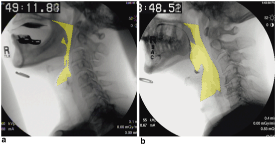
Fig. 10.1
Lateral fluoroscopic view displaying an individual with a normal (a) and dilated (b) pharyngeal area (yellow highlighted region)
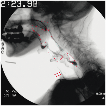
Fig. 10.2
Lateral fluoroscopic view displaying a dilated pharynx at rest (red dotted line) and aspirated barium (red arrows)
Evaluation of PES Opening
After the assessment of laryngohyoid elevation and pharyngeal contractility, the systematic assessment proceeds with the determination of the opening dimensions of the PES. The PES is a 3–4-cm-long region of high pressure. The narrowest cross section of this region is referred to as “PES opening” and is measured during passage of the largest swallowed liquid barium bolus in the lateral and AP fluoroscopic views (Figs. 10.3 and 10.4). This measurement represents the area of greatest constraint to bolus flow. If lingual or pharyngeal weakness or diminished airway protection restrict the amount of barium that can be safely administered to the patient, the assessment of accurate PES opening will be limited. Normative data for objective PES opening are influenced by age, gender, and bolus size and are available for young and elderly (> 65 years) male and female persons for both 10 and 20 cc boluses (Chap. 7). Normal mean PES opening with a 20 cc bolus is 0.90 cm (+/−0.28) for a person < 65 and 0.80 cm (+/−0.20) for an individual > 65 years of age. Diminished PES opening can be secondary to diminished elevation, poor lingual and pharyngeal constriction, decreased compliance as occurs with radiation induced stenosis, cricopharyngeus muscle (CPM) dysfunction, web, and neoplasm. After the assessment of PES opening, the systematic evaluation of the PES proceeds with an assessment of the posterior cricoid (PC) region .
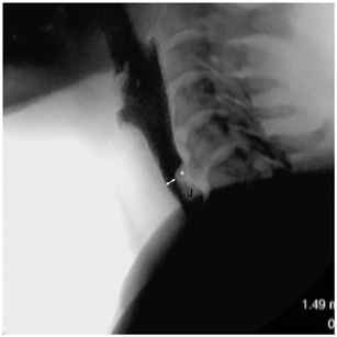
Fig. 10.3
Lateral fluoroscopic view displaying region of PES opening measurement (double white arrow). Also visible is a cricopharyngeus muscle bar (asterisk) and a jet effect (J) below the bar. A jet is a pocket of air that is created from the flow of barium beneath a region of obstruction
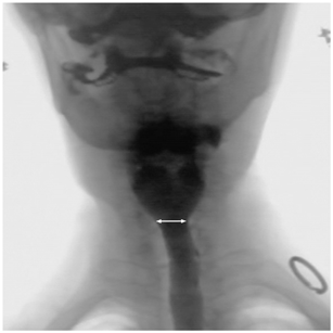
Fig. 10.4
Anterior-posterior fluoroscopic view displaying region of PES-opening measurement (double white arrow)
Evaluation of the PC Region
The PC region is defined as the area immediately adjacent to the posterior rim of the cricoid cartilage on the anterior wall of the esophageal inlet. It is differentiated from the posterior hypopharyngeal region, which is the adjacent region on the dorsal side of the esophageal inlet (Fig. 5.11). Findings of the PC region include cricoid impressions, plications, and webs. Findings of the posterior hypopharyngeal region include CPM bars and diverticuli . Precise fluoroscopic evaluation of these regions requires a large (20 cc) bolus to adequately distend the larynx and cricoid off of the spine. The inability to consume a large bolus due to lingual or pharyngeal weakness or unsafe airway protection will have a significant impact on the ability to identify PES opening and pathology.
Although not pathologic, the presence of a PC plication or PC impression is noted (Figs. 5.12 and 5.13). Cricopharyngeal webs are pathologic and are present in 14 % of dysphagic persons undergoing a VFSS. Large webs are readily visible and can tether the CPM and give the appearance of a CPM bar (Figs. 10.5 and 10.6) . It is essential to identify a web that is tethering the CPM because treatment with simple dilation is usually curative and a more invasive CPM myotomy is unnecessary. Small webs do not tether the CPM and are easily missed. They can only be visualized with the administration of a large barium bolus, a high index of suspicion, and stop-motion analysis of video with a minimum capture rate of 30 frames per second (fps) (Figs. 5.14, 10.7) . The addition of a 13 mm barium tablet to the VFSS helps identify webs that reduce the pharyngoesophageal lumen to 13 mm or less but does not assist with the recognition of smaller webs. Small cricopharyngeal webs are one of the most commonly missed fluoroscopic abnormalities in persons with solid food dysphagia . After the PC region is assessed, attention is then turned towards pathology of the posterior hypopharyngeal region.
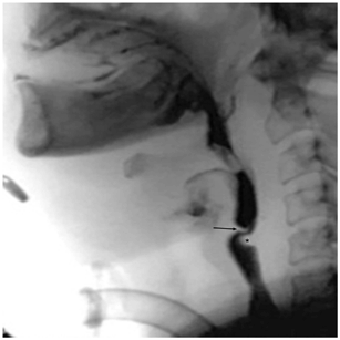
Fig. 10.5
Cricopharyngeal web on lateral fluoroscopic view. The web is protruding off of the posterior rim of the cricoid cartilage (black arrow). The cricopharyngeus muscle (asterisk) is tethered to the web severely reducing opening of the upper esophageal sphincter
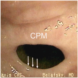
Fig. 10.6
Cricopharyngeal web visualized on endoscopy. The web (white arrows) is tethering the cricopharyngeus muscle (CPM), severely reducing opening of the upper esophageal sphincter. Cricopharyngeal webs are not usually visible on endoscopy. This view was obtained by stop-motion frame-by-frame analysis during an eructation
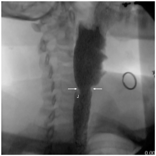
Fig. 10.7
Small cricopharyngeal web (white arrows) on right anterior oblique view. There is a small jet (J) effect
Evaluation of the Posterior Hypopharyngeal Wall Region
The most common abnormality of the posterior hypopharyngeal region is the presence of a CPM bar . CPM bars are due to a hypertrophied or poorly relaxing CPM and are not necessarily pathologic. They are seen in 30 % of normal elderly individuals without dysphagia. We differentiate CPM bars into non-obstructing (NOB) , moderately obstructing (MOB) , and severely obstructing bars (SOB) based on the degree of minimum PES opening observed with a 20 cc bolus. A NOB (Fig. 10.8) is defined as the presence of a CPM bar with normal PES opening (> 0.60 cm). A MOB is defined as the presence of a CPM bar with PES opening between 0.30 and 0.60 cm (Fig 10.9) and a SOB exists when PES opening is < 0.30 cm (Fig. 10.10) . Dysphagia is a symptom and dependent on patient-specific factors such as dentition, diet, chewing habits, and degree of visceral sensitivity. We have encountered patients with NOBs who experience significant dysphagia and patients with SOBs who experience little if any dysphagia. Association of objective findings on VFSS with subjective symptoms as documented on the Eating Assessment Tool (EAT-10) is necessary to formulate a patient-specific treatment strategy.
Stay updated, free articles. Join our Telegram channel

Full access? Get Clinical Tree


