Abdominal Aortic Coarctation and Hypoplasia


Abdominal Aortic Coarctation and Hypoplasia


James C. Stanley, Dawn M. Coleman and Jonathan L. Eliason


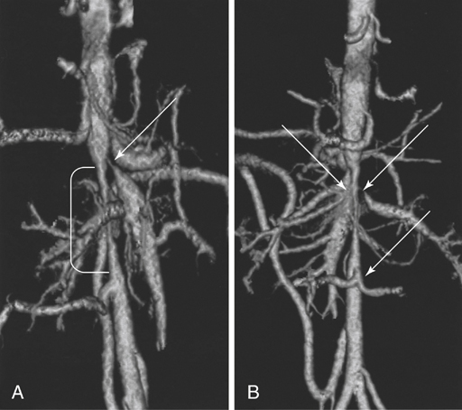
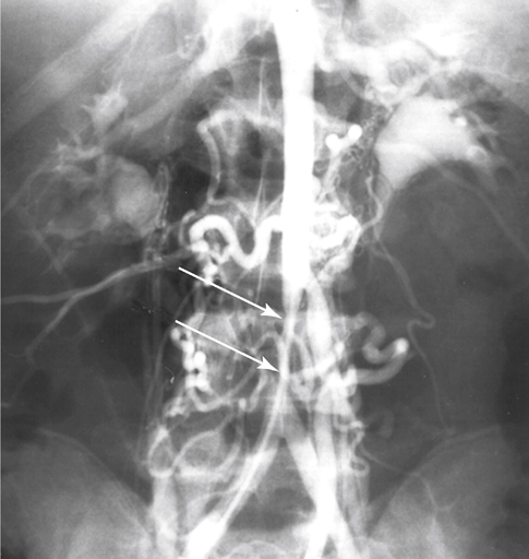
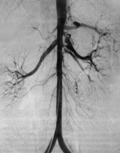
Focal developmental narrowings of the distal thoracic and abdominal aorta are rare anomalies accounting for less than 2% of all aortic coarctations. Most physicians have limited experience with the management of these unusual lesions. Abdominal aortic coarctations are categorized by the most superior extent of the aortic narrowing. Among these aortic coarctations, 69% are suprarenal (Figure 1). Narrowings originating in the intrarenal aorta occur in 23% of patients (Figure 2) and in the infrarenal aorta in 8% of patients (Figure 3). Diffuse involvement of the entire abdominal aorta is usually classified in the suprarenal category (Figure 4).


Aortoiliac hypoplasia occurs in a distinct subgroup of patients, mostly younger women, with atherosclerosis often affecting the narrowed aorta and iliac arteries. In fact, approximately 2% of women with arteriosclerotic aortoiliac occlusive disease manifest this entity. Regardless of the particular nomenclature used, most evidence suggests that both aortic coarctation and aortoiliac hypoplasia represent a spectrum of developmental abnormalities. Variations in their clinical presentation relate more to different anatomic locations and secondary pathologic events than the underlying etiologic differences. An exception exists with aortic narrowings caused by an inflammatory aortoarteritis such as occurs in Takayasu’s syndrome, in which other systemic manifestations may be more prominent than the vascular symptoms.


Etiology


The most widely accepted hypothesis as to the cause of the developmental narrowings relates to abnormal fetal events, including faulty fusion of the two primitive dorsal aortas during intrauterine life. Indirect evidence in support of this is derived from the fact that multiple renal arteries occur in 70% of patients with narrowings of the midabdominal aorta. This is two to three times the expected incidence of multiple renal arteries in the general population. The basis for this is complex.


During normal development in most embryos, fusion of the paired dorsal aortas occurs simultaneously with regression of all but one dominant metanephric vessel to each kidney around day 25 of embryonic life. It is speculated that development of a single renal artery to each kidney occurs because of its obligate hemodynamic advantage over adjacent metanephric vessels. By creating local blood flow disturbances with an aortic fusion abnormality, this obligate hemodynamic advantage is lost and additional metanephric channels persist. Support for an overfusion of the two aortas comes from the finding of a single bifurcating distal lumbar artery in up to 55% of patients with abdominal aortic coarctations and aortoiliac hypoplasia (see Figure 1), compared to less than 5% in normal persons. Multiple renal arteries are less likely to develop when the aortic coarctation is distant from the central abdominal aorta.


Theoretically, any event that alters the normal transition or organization of fetal mesenchymal cells into vascular smooth muscle can result in narrowing of the aorta as well as splanchnic and renal arterial stenoses. This might explain the existence of aortic coarctation or hypoplasia following cytocidal viral infections such as gestational rubella. A genetic alteration in the growth and development of vascular smooth muscle has also been implicated in abdominal aortic coarctation and hypoplasia associated with neurofibromatosis 1 and Williams syndrome. Coincident with the aortic narrowings in patients with Williams syndrome are the common occurrence of renal and splanchnic arterial narrowings. These arteries at their aortic origins are truly diminutive. It is more than happenstance that among our patients with midabdominal aortic narrowings, renal arterial stenoses were seen in 87% and splanchnic arterial occlusive disease in 62%.


Clinical Manifestations


Developmental narrowings of the distal thoracic and midabdominal aorta generally become evident during the first or second decade of life, with a mean age at initial diagnosis of 12 years. There appears to be a slight male gender predilection. The classic triad in these young patients consists of severe hypertension, diminished or absent femoral pulses, and an abdominal bruit. Symptomatic lower extremity ischemia and intestinal angina affect less than 10% of these children.


The natural history of patients with developmental narrowings of the more cephalic abdominal aorta above the renal vessels is directly related to the severity of their renin-mediated hypertensive vascular disease. The prognosis for untreated patients is dismal, and most patients die in early adulthood from cardiac failure or cerebrovascular accidents. In one earlier review, 55% of untreated patients died at a mean age of 34 years. Thus, all patients with coarctation or segmental hypoplasia of the distal thoracic or central abdominal aorta must be considered at risk for serious complications of their disease. Infrarenal abdominal aortic narrowings, unlike those above the renal arteries, usually become symptomatic in the fourth or fifth decade of life as a result of secondary atherosclerotic occlusive disease. These patients often come to the hospital with lower extremity fatigue and less often with claudication.


There is a generally recognized predilection for women to exhibit hypoplastic aortoiliac narrowings. Although the genetic basis for this phenomenon is unknown, anecdotal evidence suggests a higher incidence among women with red hair. Their usual presentation is lower extremity claudication.

Stay updated, free articles. Join our Telegram channel

Aug 25, 2016 | Posted by in CARDIOLOGY | Comments Off on Abdominal Aortic Coarctation and Hypoplasia

Full access? Get Clinical Tree

Get Clinical Tree app for offline access