3D Echo
Dr Thomas Mathew
Trent Cardiac Centre, Nottingham, UK
The heart is a three-dimensional (3D) structure and so the display of anatomy in 3D makes assessment easier and more accurate. Using two-dimensional (2D) echo, this is usually achieved by the examiner mentally reconstructing a 3D image from the information obtained from multiple 2D imaging planes. Three-dimensional echo allows the acquisition of 3D volume datasets and the instantaneous real-time display of a beating heart in three dimensions. This is a major breakthrough in the field of echocardiography.
3D TECHNOLOGY
The backbone of 3D technology is the transducer. Current systems use fully-sampled matrix array transducers. A 2D transducer typically contains 128 elements arranged in a linear fashion. The ultrasound beam is steered across one plane (y axis, azimuth plane) creating a tomographic slice of the heart. In contrast, matrix array transducers contain nearly 3000 elements arranged in the form of a rectangular grid and capable of parallel processing. The ultrasound beam can be steered in two different planes – the y axis (similar to 2D imaging) and, in addition, the z axis (elevation plane) – to produce a pyramidal volume dataset (Fig. 12.1).
Because of the large number of elements, some form of processing (for example, beam forming) has to be done within the transducer itself. This is to avoid having to connect every element to the ultrasound machine, which would make the cabling impossibly heavy. For this purpose, miniaturised circuit boards are incorporated within the transducer. As a consequence, 3D transducers in general are larger and heavier than the conventional 2D transducers. However refinements in transducer technology have enabled the most recent generation of matrix array transducers (e.g. X5-1 from Philips Medical Systems) to be significantly smaller in size.
In addition to transducer design, advances in computer processing power and the availability of software packages for offline and online analysis have allowed 3D echo to become a practical clinical tool. Currently 3D imaging is available for both transthoracic and transoesophageal examinations.
3D echo physics
Similar to 2D imaging, there is an inverse relationship between frame rate (temporal resolution), sector width and spatial resolution (scan lines) in 3D echo. An increase in one of these factors will cause a decrease in the other two. Manufacturers have developed several techniques such as parallel processing and multibeat imaging to cope with this challenge but in practice this is usually achieved by selecting the appropriate acquisition mode or by using a small volume dataset. In this way a diagnostic quality image can be obtained with sufficient spatial resolution and temporal resolution.
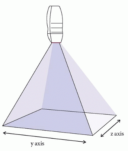 Figure 12.1 The ultrasound beam from a 3D matrix array transducer is steered in two different planes to create a pyramidal volume dataset |
Acquisition modes
There are two methods of data acquisition in 3D echo:
real-time or live 3D imaging
multi-beat imaging.
Real-time (live) 3D imaging acquires a pyramidal volume dataset from one cardiac cycle and projects the image as an online real-time display. As the information is updated in real time, image orientation and plane can be changed by rotating the transducer. Analysis can be done with minimal post-processing and the image display can be rotated (independent of the transducer position) to view the heart from different orientations. Whilst this method is useful to assess the real-time motion of a cardiac structure, it is limited by spatial and temporal resolution. Realtime imaging is available in the following modes:
Live 3D: Any 2D image can be converted to a 3D image by single-button activation in this mode. To allow sufficient spatial and temporal resolution, the software automatically defaults to a narrow sector width of approximately 300 × 600 pyramidal volume (Fig. 12.2). The sector width can be increased to
visualise a larger structure, but the scan density (spatial resolution) and frame rate (temporal resolution) will go down.

Figure 12.2 Live 3D image (right-hand panel) of a parasternal long axis view obtained from a single beat
Live 3D colour: Colour flow can be superimposed on a live 3D image to assess blood flow in real time. This facility is only available from some manufacturers and with the most recent software update.
3D zoom: This is an extension of live 3D that allows a focussed real-time view of a structure of interest. By adjusting the lateral and elevation width using a crop box placed on a pre-acquired image, the system automatically crops the adjacent structures to provide a real-time display of the structure of interest (Fig. 12.3).
Multiplane imaging: One unique feature of the matrix array transducer is to display 2D images in multiple planes. For example, acquisition of a 4-chamber view from the apical window will simultaneously display 2-chamber and 3-chamber views (Fig. 12.4). The first image is often the reference image and the
others can be changed depending on the operator chosen angle. Although strictly not a 3D image, this feature is useful in situations where assessment of multiple imaging planes from the same cardiac cycle is useful (e.g. stress echo, tissue synchronisation imaging).
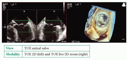 |
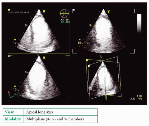 Figure 12.4 Multiplane imaging. Simultaneous display of 4-, 2- and 3-chamber (apical long axis) views from a single acquisition from the apex. Contrast has been administered to improve endocardial definition |
Multi-beat imaging, in contrast to real-time/live 3D imaging, acquires several small volume real-time datasets from four to six cardiac cycles and stitches them together to create an instantaneous full volume display. As the images are acquired over several cardiac cycles, it is ‘near real-time’ than real-time. Once acquired, the images cannot be altered by manipulation of the probe as in live 3D


Stay updated, free articles. Join our Telegram channel
Full access? Get Clinical Tree
