Fig. 1.1
Anterior view of sternum with clavicular and costal cartilage attachments. Surface landmarks denoted (suprasternal notch and sternal angle)
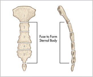
During development, the sternum consists of six sternebrae (Fig. 1.2). During late adolescence, the central four segments fuse to form the sternal body . The sternal body is bordered bilaterally by the articulation of the second through seventh costal cartilages. A common variant in females is the joining of the facets for the sixth and seventh costal cartilages. The lateral sternal border also attaches to the internal intercostal muscles and anterior intercostal membrane while the pectoralis major muscle attaches along the anterior–lateral surface.


Fig. 1.2
Developing sternum which consists of six separate sternabrae. The central four segments fuse during adolescence
The xiphoid process is a cartilaginous structure that attaches inferiorly to the sternal body along with connections to the costal margin of the seventh rib via the costoxiphoid ligament and a direct attachment to the rectus sheath. It is noted to be variable in length and structure with the most common variants being bifid or perforated. The xiphoid process is occasionally prominent enough to be seen on physical exam as a protrusion at the inferior border of the sternum.
Ribs
The thoracic rib cage is a diverse structure built for security and support of the underlying organs but is uniquely designed to facilitate respiration. There are 12 paired ribs along with their associated costal cartilages that develop from the costal process of each individual thoracic vertebrae. During development, rib pairs migrate superiorly so that they not only abut their original vertebra but also the one immediately cephalad. This migration affects ribs 2 through 9 and occasionally the tenth rib. The cephalic migration of the vertebral end relative to the sternal end of the ribs explains the characteristic shape of the rib cage.
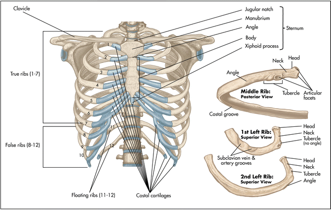
Ribs 1 through 7, referred to as true ribs , articulate with the manubrium or sternum while ribs 8 through 10, known as false ribs, articulate with the costal cartilage of the adjacent ribs. Ribs 11 and 12 do not have an anterior articulation point and are therefore free floating (Fig. 1.3).


Fig. 1.3
Anterior view of the bony thorax and pectoral girdle. True, false and floating ribs are denoted. Insert contains images of a typical rib and the first rib
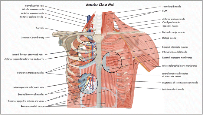
Basic rib anatomy consists of a head, neck, tubercle, angle, shaft, and costal groove. The head of a typical rib articulates at two points, the superior costal facet of the thoracic vertebra of the same number and the inferior costal facet of the thoracic vertebra just cephalad. An articular capsule surrounds the head of each rib and is further attached to the vertebra via the radiate ligament. The tubercule articulates with the transverse process of the adjacent vertebra along with the presence of multiple costotransverse ligaments for added support. As you follow the contour of the bony rib to the end of the shaft, you reach the costochondral joint where the bony rib and cartilage are securely attached via the intertwining of periosteum and perichondrium. The attachment of the first ribs is through synchondroses or cartilaginous joints and are therefore immobile while the second through seventh rib pairs articulate with the sternum via synovial joints that allow for movement during respiration. These joints are reinforced by sternocostal ligaments. The costal groove lies along the inferior surface and houses the intercostal neurovascular bundle (Fig. 1.4).


Fig. 1.4
Anterior chest wall showing muscular attachments and neurovascular structures
Ribs 3 through 9 are typical ribs as described earlier while ribs 1, 2, 10, 11, and 12 are atypical. The first rib is a short, flat rib that is much wider and more curved than those previously described. Its head has only one facet for articulation with the body of the first thoracic vertebrae and the prominent tubercle forms a synovial joint with the transverse process. Anteriorly, the first rib is secured to the manubrium via a synchondrosis and the clavicle by the costoclavicular ligament. It has attachments from the anterior, middle, and posterior scalene muscles as well as the serratus anterior, subclavius, and erector spinae muscles. The subclavian artery and vein cross over the first ribs near the middle of the shaft and under the clavicles bilaterally. The second rib is unique in that it has a large tuberosity for attachment of the serratus anterior muscle and a poorly developed costal groove. The head of ribs 10–12 articulates with the corresponding thoracic vertebrae via one facet. Ribs 11 and 12 are further distinguished by lacking a neck, angle, and costal groove. They also do not articulate with the corresponding transverse process.
Stay updated, free articles. Join our Telegram channel

Full access? Get Clinical Tree


