Lung, Chest Wall, Pleura, and Mediastinum

Chapter 58 Lung, Chest Wall, Pleura, and Mediastinum


The term thorax refers to the area between the neck and abdomen enclosed by the ribs, sternum and vertebrae radially, the thoracic inlet superiorly, and the diaphragm inferiorly. The chest or thorax supports and protects the internal thoracic organs, provides for the negative inspiratory force that initiates ventilation and the positive expiratory force needed for vocalization, and creates a frame for the neck, upper extremities, thoracic structures, and abdomen. The major thoracic structures include the heart and lungs, chest wall, including the overlying musculature, ribs, sternum, vertebrae, diaphragm, trachea, and great vessels.


Anatomy


The thoracic organs are protected by the bony thorax and overlying chest musculature. The parietal pleura, the internal lining of the chest wall, is separated from the visceral pleura, the outer lining of the lung, by a small amount of pleural fluid. The parietal pleura covers the chest wall, mediastinum, diaphragm, and pericardium. The visceral pleura covers the lung and separates the lobes from one another. The pleural space is a potential space that may compress the lungs or heart with fluid, tumor, or infection. The right and left pleural spaces are separated from one another by the mediastinum.


The bony thorax is covered by three groups of muscles—the primary and secondary muscles for respiration and those attaching the upper extremity to the body (Fig. 58-1). The primary muscles include the diaphragm and intercostal muscles. The intercostal muscles of the intercostal spaces include the external, internal, and transverse or innermost muscles. Eleven intercostal spaces, each associated numerically with the rib superior to it, contain the intercostal bundles (vein, artery, and nerve) that travel along the lower edge of each rib. All intercostal spaces are wider anteriorly and each intercostal bundle falls away from the rib posteriorly to become more centrally located within each space. The intercostal muscle layers assist with respiration and protect the thoracic structures. The extrinsic muscles of the chest, latissimus dorsi muscle, serratus anterior muscle, pectoralis major and minor muscles, and cervical muscles (sternocleidomastoid, scalene muscles) attach to the bony thorax, protect the chest wall itself, and may assist with ventilatory efforts in those with chronic obstructive pulmonary disease (COPD).
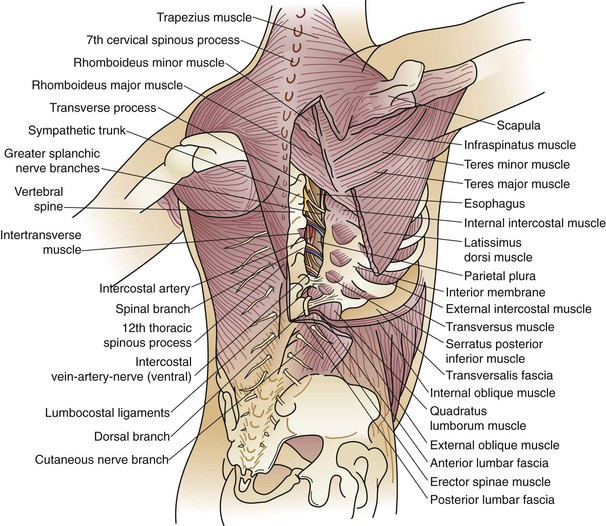


The secondary muscles consist of the sternocleidomastoid, serratus posterior, and levatores costarum. The third muscle group attaches the upper extremity to the body. The pectoralis major and minor muscles lie anteriorly and superficially. Posterior superficial musculature includes the trapezius and latissimus dorsi. Deep muscles include the serratus anterior and posterior, levatores, and major and minor rhomboids. These superficial and deep muscles help hold the scapulae to the chest wall. In respiratory distress, the deltoid, pectoralis, and latissimus dorsi muscles form a tertiary system for ventilatory assistance through fixation of the upper extremities.


The bony thorax consists of 12 ribs peripherally extending from the vertebrae posteromedially, to the sternum or costal arch anteriorly (Fig. 58-2). The 11th and 12th ribs are floating ribs and are not attached directly to the sternum. Ribs 1 to 5 are directly attached to the sternum by costal cartilages. The lower ribs (6 to 10) coalesce into the costal arch. The first rib is relatively flat, dense, and travels from the first thoracic vertebra to the manubrium to create the thoracic inlet (Fig. 58-3). Through this relatively small area pass the great vessels, trachea, esophagus, and innervation to the upper extremity, diaphragm, and larynx. Trauma to this area, manifested by a first rib fracture, is the consequence of a significant mechanical force with likelihood of injury to one or more of these structures. Other structures within the thoracic inlet include the phrenic nerve, recurrent laryngeal nerve in the tracheoesophageal groove, which recurs around the aorta at the ligamentum arteriosum on the left and around the innominate artery on the right, and insertion of the thoracic duct posteriorly at the junction of the left subclavian with the left internal jugular veins. The remaining ribs gradually slope downward. Each rib is composed of a head, neck, and shaft. Each head has an upper facet, which articulates with the vertebral body above it, and a lower facet, which articulates with the corresponding thoracic vertebra to that rib, establishing the costovertebral joint. The neck of the rib has a tubercle with an articular facet; this articulates with the transverse process, creating the costotransverse joint and imparting strength to the posterior rib cage.
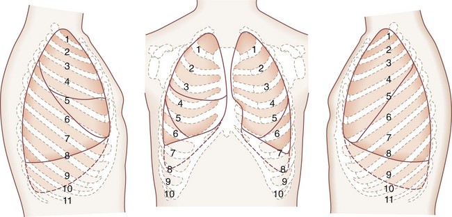
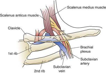


The sternum is flat, 15 to 20 cm in length, approximately 1.0 to 1.5 cm in thickness, and comprised of the manubrium, body, and xiphoid. The manubrium articulates with each clavicle and the first rib. The manubrium joins the body of the sternum at the angle of Louis, which corresponds to the anterior aspect of the junction of the second rib. The angle of Louis is a superficial anatomic landmark for the level of the carina. The anterior cartilaginous attachments of the true ribs to the sternum, along with intercostal muscles and the hemidiaphragms, allow for movement of the ribs with respiration.


The trachea in adults is approximately 12 cm in length, with 18 to 22 cartilaginous rings. The internal diameter is 2.3 cm laterally and 1.8 cm anteroposteriorly. The larynx ends with the inferior edge of cricoid cartilage. The cricoid is the only complete cartilaginous ring in the trachea. The trachea begins approximately 1.5 cm below the vocal cords and is not rigidly fixed to surrounding tissues. Vertical movement is easily possible. The most rigid point of fixation is where the aortic arch forms a sling over the left mainstem bronchus. The innominate artery crosses over the anterior trachea in a left inferolateral to high right anterolateral direction. The azygos vein arches over the proximal right mainstem bronchus as it travels from posterior to anterior to empty into the superior vena cava. The esophagus is closely applied to the membranous trachea and lies to the left of the midline of the trachea. The recurrent laryngeal nerves run in the tracheoesophageal groove on both the right and left. The blood supply to the trachea is lateral and segmental from the inferior thyroid, internal thoracic, supreme intercostal, and bronchial arteries. Circumferential dissection more than 1 to 2 cm during tracheal reconstruction may lead to vascular insufficiency, with necrosis or anastomotic dehiscence.


Lung development begins at approximately 21 to 28 days’ gestation. The true alveolar stage, with air sacs surrounded on all sides by capillaries, occurs from approximately 7 months to term. Alveolar proliferation continues after birth. There are approximately 20 million alveoli at birth, which increase to approximately 300 million by age 10 years, with no more increase after that time. There are 23 generations of bronchi between the trachea and terminal alveoli. In the lung, 80% of its volume is air, 10% is blood, and approximately 10% is solid tissue. Alveoli make up approximately 50% of the entire lung volume.


The lungs are broadly divided into five lobes, with multiple segments in each lobe (Fig. 58-4). The right lung is composed of three lobes, the upper, middle, and lower. Two fissures separate these lobes. The major, or oblique, fissure separates the lower lobe from the upper and middle lobes. The minor or horizontal fissure separates the upper lobe from the middle lobe. The left lung has two lobes—the upper lobe and lower lobe. The lingula corresponds embryologically to the right middle lobe. A single oblique fissure separates the lobes.
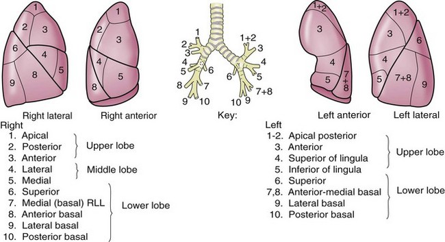


The bronchopulmonary segments are divisions of each lobe that contain anatomically separate arterial, venous, and bronchial supplies. There are 10 bronchopulmonary segments on the right and eight bronchopulmonary segments on the left.


The blood supply of the lung is twofold. Unoxygenated blood circulates from the right ventricle through the pulmonary artery to each lung. After oxygenation in the lung, the blood is returned to the left atrium through the pulmonary veins. Blood supply to the bronchi is from the systemic circulation via bronchial arteries arising from the superior thoracic aorta or the aortic arch, either as discrete branches or in combination with the intercostal arteries.


Lymphatic vessels are present throughout the lung parenchyma and pleura and gradually coalesce toward the hilar areas of the lungs. Generally, lymphatic drainage from the lung affects the ipsilateral lymph nodes; however, flow of lymph from the left lower lobe may drain to the right mediastinal (paratracheal) lymph nodes. Lymphatic drainage within the mediastinum moves cephalad. The pulmonary parenchyma does not contain a nerve supply.


The visceral pleura is separated from the parietal pleura by a small amount of pleural fluid that allows almost frictionless movement during respiration. The blood supply of the parietal pleura comes from the systemic arteries and veins, including the posterior intercostal, internal mammary, anterior mediastinal, and superior phrenic arteries, and corresponding systemic veins. The blood supply of the visceral pleura is systemic and pulmonary. The lymphatic drainage of the parietal pleura is into regional lymph nodes, including the intercostal, mediastinal, and phrenic nodes. Visceral pleural lymphatics follow the superficial lung lymphatics and drain into the mediastinal lymph nodes. The parietal pleura underlying the ribs has rich nerve endings from the intercostal nerves. Generous local anesthesia is therefore necessary for chest tube insertion. The visceral pleura is innervated by vagal branches and the sympathetic system.


The anatomic boundaries of the mediastinum include the thoracic inlet superiorly, diaphragm inferiorly, sternum anteriorly, vertebral column posteriorly, and medially to the parietal pleura. Thoracic tumors that penetrate through the pleura (by definition) invade the mediastinum. Traditionally, the mediastinum can be divided into anterosuperior, middle, and posterior compartments. There no specific anatomic planes that define these areas. Fat and lymph nodes are found throughout the mediastinum.


The anterosuperior compartment includes the thymus gland. The right and left lobes of the thymus extend into the cervical areas; these portions of the thymus must be resected to provide for complete extirpation of the gland.


The middle mediastinum contains the heart, pericardium, great vessels, including the descending, transverse, and descending aorta, superior and inferior vena cava, pulmonary artery and veins, trachea and bronchi, and phrenic, vagus, and recurrent laryngeal nerves. The phrenic nerve enters the thorax through the thoracic inlet on the anterior aspect of the anterior scalene muscle.


The vagus nerve enters the thoracic inlet through the carotid sheath. It lies anterior to the subclavian and posterior to the innominate artery on the right. The right recurrent laryngeal nerve loops or recurs around the innominate artery to innervate the right vocal cord. The vagus nerve then continues posteriorly in the tracheoesophageal groove to innervate the trachea and continues down to innervate the esophagus. On the left side, the vagus nerves enters the thorax through the thoracic inlet and, as it exits the carotid sheath, moves along the anterior aspect of the aortic arch. The recurrent laryngeal nerve arises from the vagus nerve, loops around under the ligamentum arteriosum, continues superiorly under the aorta, and lies in the tracheoesophageal groove as it innervates the left recurrent laryngeal nerve. The left vagus continues posteriorly within the mediastinum posteriorly along the esophagus to innervate the trachea and esophagus.


The posterior mediastinum contains those structures between the heart and pericardium and trachea anteriorly, and the vertebral column and paravertebral spaces posteriorly. The posterior mediastinum contains the esophagus, descending aorta, azygos and hemiazygos veins, thoracic duct, sympathetic chain, and lymph nodes. The thoracic duct originates from the cisterna chyli in the abdomen. It enters the chest through the aortic hiatus in an anterolateral position, and travels superiorly just to the right of midline in the chest along the anterolateral surface of the vertebral column. At approximately the level of T5, it crosses over to the left and continues superiorly to empty, posteriorly, into the junction of the left jugular and subclavian veins.


The inferior border of the mediastinum is the diaphragm, which separates the abdominal contents from the thorax. Hernias through the esophageal hiatus (paraesophageal hernias), or through the foramen of Bochdalek (posteriorly) or the foramen of Morgagni (anteriorly), may be initially identified as a mediastinal mass.


Each spinal root exits the neural foramina of the vertebral body and bifurcates to form a branch to the intercostal nerve, to innervate the skin and intercostal musculature, and a branch to the sympathetic ganglion. Intercostal nerves innervate the skin and musculature of the intercostal muscles. The spinal root divides as it exits the neural foramina. One branch goes to the intercostal nerve and one lies in the posterior vertebral gutter to form the sympathetic ganglion. The thoracic sympathetic trunk is composed of several ganglia that lie along the ribs. The most superior ganglion is the stellate ganglion.


Selection Of Patients For Thoracic Operations


The physiologic evaluation of the thoracic surgical patient must be individualized for each patient, but generally emphasizes pulmonary and cardiac function. The assessment of a patient’s ability to tolerate lung resection from a cardiopulmonary standpoint is fundamental to patient selection for surgery. Patients with advanced pulmonary disease and severe pulmonary dysfunction may have a prohibitive risk, which may exist in more than one third of patients with otherwise resectable lung disease.1


Cigarette smoking is associated with up to a sixfold increase in the incidence of postoperative pulmonary complications after surgery.2 If the patient is a smoker, he or she must stop smoking immediately. The physician must clearly communicate this message. Although there are few studies specific to pulmonary resection, there is evidence that preoperative smoking abstinence of 4 to 8 weeks’ duration is necessary to reduce the incidence of complications. Ideally, patients are smoke-free for a minimum of 2 weeks and preferably 4 to 8 weeks before surgery,3 although smoking cessation at any time is valuable.4 Smoking cessation programs may be helpful for these patients, and they may need pharmacologic assistance. This combination may have increased efficacy in smoking cessation efforts over counseling alone.


Prior to the operation, and in the perioperative period, deep venous thrombosis prophylaxis is provided by subcutaneous heparin and/or by sequential compression stockings. Also, perioperative antibiotics are used to minimize complications from infections. Postoperative morbidity may also be minimized by adequate pain control to facilitate early ambulation. Routine use of a thoracic epidural catheter (or patient-controlled analgesia [PCA]) provides excellent pain control. Incentive spirometry assists in expanding the lung and reducing the incidence of pulmonary morbidity. Nasal bilevel positive airway pressure for patients with obstructive sleep apnea may delay or eliminate the need for intubation or reintubation after pulmonary resection. Early mobilization is essential to avoid most perioperative complications.


Physiologic Evaluation


Before thoracic operations, patients may be evaluated by a combination of physiologic studies.5 A plain chest roentgenogram is commonly obtained (Fig. 58-5). Spirometry measures the lung volumes (Fig. 58-6) and mechanical properties of lung elasticity, recoil, and compliance. Pulmonary function testing (Fig. 58-7) also evaluates gas exchange functions, such as DLCO (diffusion of carbon monoxide in the lung).


The predicted postoperative forced expiratory volume (FEV) in 1 second (FEV1) is the most commonly used indicator of postoperative pulmonary reserve. Most patients with an FEV1 in excess of 60% predicted will tolerate an anatomic lobectomy, depending on other evaluable factors. If the FEV1 is less than 60% of predicted, further testing might be considered in an attempt to estimate postoperative FEV1 (predicted postoperative FEV1 [ppo-FEV1]). The quantitative ventilation-perfusion lung scan is used to assist in the calculation of postoperative residual pulmonary function after resection. Patients with a ppo-FEV1 of 35% to 40% should functionally tolerate the operation.


Quantitative radionucleotide perfusion scanning involves the injection of 99mTc-radiolabeled albumin particles followed by the visual inspection of planar images (Fig. 58-8). Quantitative perfusion provides a measurement of the relative function of each lobe and lung, allowing a prediction of pulmonary function after lung resection:


image


A postoperative FEV1 less than 30% predicted carries a greater postoperative risk for oxygen, and even ventilator dependence, but a decision to deny surgical resection to this group of patients must be considered on an individual basis because some will do better than expected with careful selection at experienced centers. Finally, in the immediate postoperative period, the objectively calculated ppo-FEV1 will likely not be realized secondary to limited ambulation, pain, or other emotional or physical factors.


The carbon monoxide diffusing capacity (DLCO) can be measured by several methods, although the single-breath test is most commonly performed. The DLCO measures the rate at which test molecules such as carbon monoxide move from the alveolar space to combine with hemoglobin in the red blood cells. The DLCO is determined by calculating the difference between inspired and expired samples of gas. DLCO levels less than 40% to 50% are associated with increased perioperative risk.6


The ratio of FEV1 in 1 second to forced vital capacity ratio (FEV1/FVC) describes the relationship between the FEV1 and the functional lung volume. In obstructive disease, the ratio is low (FEV1 is low and FVC is high); in restrictive disease, the ratio is approximately normal because both FEV1 and FVC are reduced.


Flow-volume loops derived from spirometry describe the relationship between lung volume and air flow as the lung volume changes during a forced expiration and inspiration. The typical test consists of tidal breathing at rest, maximal inspiratory effort to total lung capacity, and maximal expiratory effort to residual volume, concluding with maximal inspiratory effort to total lung capacity.


Cardiopulmonary Exercise Testing


Cardiopulmonary exercise testing (CPET) can be extremely useful for the evaluation of marginal candidates (ppo-FEV1 or ppo-DLCO <50% predicted) or for patients who appear more disabled than expected from simple spirometry measurements. Formal CPET includes an exercise electrocardiogram (ECG), heart rate response to exercise, and measurements of minute ventilation and oxygen uptake/min. CPET allows a calculation of maximum oxygen consumption (image) and provides insight into overall cardiopulmonary function (the cardiopulmonary axis) that cannot be ascertained from other objective studies. CPET may identify clinically occult cardiac disease and provide a more accurate assessment of pulmonary function than spirometry and DLCO, which tend to overestimate functional loss after resection.


A patient’s risk of perioperative morbidity and mortality may be stratified by image. Those with image above 20 mL/kg/min are not at increased risk for complications or death after resection of non–small cell lung cancer (NSCLC). A level below 15 mL/kg/min is associated with an increased risk, and image less than 10 mL/kg/min indicates very high risk, generally precluding operation.7,8 Some have advocated stair climbing as a suitable measure of preoperative cardiopulmonary assessment.9 Given the wide availability of more objective and standardized noninvasive tests for cardiopulmonary function, stair climbing performance should not be used as the sole criterion to determine physiologic suitability for lung cancer resection. In patients undergoing evaluation for lung volume reduction surgery or for lung transplantation, a 6-minute walk test is used for a measure of the cardiac and pulmonary reserve. Patients are told to walk as far and as fast as they can during this time period. Distances of more than 1000 feet suggest an uncomplicated course.


Measurement of diaphragm function by fluoroscopy, the sniff test, is needed to determine symmetry of effort and exclude paradoxical movement of the diaphragm. Paradoxical movement—elevation of one hemidiaphragm with active contraction or retraction of the other diaphragm—suggests paresis or paralysis. This finding may suggest a specific reason for breathlessness. Diaphragm plication may be therapeutic.


No single test result should be viewed as an absolute contraindication to surgical resection. Although the physiologic assessment for patients undergoing normal spirometry and minimal comorbidity is fairly straightforward, patients with marginal preoperative indices must be considered on an individual basis.


Thoracic Incisions


The choice of incision depends on the operation, patient’s underlying physiologic condition, and anticipated benefits and limitations of the planned approach.


Video-assisted thoracic surgery (VATS) and other minimally invasive techniques have been developed to treat most thoracic problems, including lung cancer, mediastinal tumors, pleural diseases, and parenchymal diseases, and diagnosis and staging of thoracic malignancies. Minimally invasive techniques appear to minimize pain and surgical trauma from the incisions, decrease hospitalization, and improve convalescence. Small incisions are made for the camera and other instruments, depending on the location of the tumor. The ribs are not spread. Improved lighting and optics create excellent exposure and visualization.


The posterior or posterolateral thoracotomy is used for operations on a single thorax, pulmonary resection, esophageal surgery or resection, or resection of portions of the chest wall. The patient is placed in a lateral decubitus position. An oblique incision is used posteriorly or a vertical axillary incision is made just anterior to the latissimus dorsi muscle.


The anterior or anterolateral thoracotomy is created by a curvilinear incision underneath the inferior border of the pectoralis major muscle at the inframammary fold. A median sternotomy is performed using a vertical incision from the sternal notch to the xiphoid. A sternal saw is then used to divide the sternum in the midline. With gentle retraction, the sternum can be spread approximately 8 to 10 cm to allow access to the mediastinum, heart, great vessels, and right and left thorax. The pleura can be opened on either side to explore the hemithorax. The sternum is usually closed with stainless steel wire.


The transverse sternotomy, or clamshell incision, is larger than a median sternotomy and more uncomfortable for the patient. This incision combines two anterior thoracotomy incisions in the inframammary fold with transverse division of the sternum at the fourth intercostal space. Both internal mammary arteries are ligated. This approach is ideal for accessing the right and left hilum, as well as providing additional exposure for large mediastinal tumors, bilateral hilar dissections, bilateral lung transplantation, or posterior-based metastases in both lungs.


Lung


Congenital Lesions


Various congenital lung abnormalities can occur as a consequence of disturbed embryogenesis.10 Bilateral agenesis of the lungs is fatal. Unilateral agenesis may occur more frequently on the left (≈70%) than on the right (≈30%), with more than a 2 : 1 male-to-female ratio.


Hypoplasia of the lungs may occur as a result of interference with the development of the alveolar system during the last 2 months of gestation. Bochdalek’s hernia is the most frequent cause of hypoplasia. Conditions associated with hypoplasia of the lungs include oligohydramnios, prune belly syndrome (deficiency in the abdominal musculature, genitourinary abnormalities), scimitar syndrome (abnormal pulmonary vein draining into the inferior vena cava, demonstrated as a crescent along the right heart border on a cardiac angiogram), and dextrocardia. Isolated pulmonary hypoplasia is rare.


Hyaline membrane disease (or infant respiratory distress syndrome) is frequent in premature infants (24 to 28 weeks’ gestation) and infants of diabetic mothers. At that point in gestation, infants have an immature surfactant system. Hyaline membrane disease develops in the alveoli, causing congestion and a grossly deep purple–appearing lung. Respiratory distress frequently ensues, requiring high concentrations of oxygen. Chest x-rays demonstrate a ground glass appearance from the interstitial edema. As needs for oxygen and ventilator pressure increase to counteract this interstitial edema, pneumothorax frequently occurs. Of these infants, 10% to 30% do not survive.


Cystic Lesions


Congenital cystic lesions generally occur secondary to separation of the pulmonary remnants from airway branchings. Clinically, about one third of patients are without symptoms, one third have cough, and one third have infection or, rarely, hemoptysis. Treatment may be antibiotics or, for more severe localized cases, resection. Any thoracic cystic lesion that enlarges on serial radiographs needs to be considered for resection.


A bronchogenic cyst arises from a tracheal or bronchial diverticulum (see later, “Mediastinal Cysts and Tumors”). This diverticulum becomes completely separated from the trachea and is frequently found as an asymptomatic mass on routine chest x-rays. Computed tomography (CT) of the chest demonstrates this abnormality as a homogeneous-type mass, well circumscribed, and adjacent to the trachea (Fig. 58-9). The bronchogenic cyst accounts for 10% of mediastinal masses in children and is located in the midmediastinum. Treatment consists of excision, even if the patient is asymptomatic, to confirm the diagnosis.
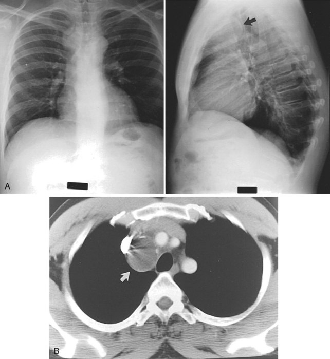


Cystic fibrosis is an autosomal recessive disorder found more commonly in whites. Approximately 20% of patients with cystic fibrosis survive to the age of 30 years. Lung failure is the most frequent cause of death in most patients. Excessively thick mucus leads to inspissation, recurrent infections, bronchitis, and bronchiectasis. Pneumothorax secondary to air trapping is also found. Fibrosis and cystic changes on pathologic examination are identified. A tension cyst may be a complication of cystic disease. A rapid increase in the size of the cyst may cause mechanical ventilation problems as well as mediastinal shift. Resection, usually lobectomy, corrects this problem. Pneumatoceles may develop as a result of childhood Staphylococcus aureus infection. They can be large and may cause mechanical complications, which may resolve completely as the pneumonia resolves. Resection may be needed.


Congenital Bronchopulmonary Malformations


Lobar emphysema10 is the most commonly resected congenital cystic lesion (50%). The onset of rapidly progressive respiratory distress usually occurs from 4 to 5 days to several weeks after birth. It rarely occurs after 6 months of age. It predominantly affects the upper lobe. Bronchiolitis is probably the most common cause overall. Treatment is lobectomy.


Congenital cystic adenomatoid malformations are the second most commonly resected congenital cystic lesion. They are closely related to a hamartoma without cartilage. Terminal bronchioles proliferate, yielding the adenomatoid malformation. The lung has the appearance of Swiss cheese and feels like a large rubbery mass. With air trapping and overdistention, respiratory distress may occur, which is optimally relieved by lobectomy.


Pulmonary sequestration is an area of embryonic lung tissue, separate from the lung, which receives blood supply from an anomalous systemic artery from the aorta, not the pulmonary artery. This condition occurs secondary to an accessory lung bud caudal to the normal lung, but with a lack of absorption of primitive surrounding splanchnic vessels. During lung development, interlobar sequestration (75%) occurs early. Later, after the pleura forms, extralobar sequestration (ELS) occurs (25%), primarily on the left side (66%), and is completely enclosed by its own pleura. The ELS blood supply is usually from the thoracic or upper abdominal aorta to systemic (azygous or hemiazygous) veins. ELS is more common in males. Resection is recommended. Intralobar sequestration (ILS) mainly occurs within the lower lobes (>95%) and is equally distributed between the right and left lower lobes. ILS blood supply is from the descending thoracic aorta, which usually traverses the pulmonary ligament. Venous drainage is via the pulmonary veins. Ninety-five percent of the systemic blood supply to the pulmonary sequestration comes from the thoracic aorta.


Congenital Abnormalities of the Trachea and Bronchi


Esophageal atresia with tracheoesophageal fistula is the most frequent abnormality of the trachea in infants (see later, “Trachea”) Bronchial atresia is the second most frequent congenital pulmonary lesion after tracheoesophageal fistula.11 The lung tissue distal to the atresia expands and becomes emphysematous as a result of air entry through the pores of Kohn. With no exit for air or mucus because of this blind bronchial stump, emphysema from air trapping or development of a mucocele may develop. Chest x-rays may demonstrate hyperinflation of a lobe or segment. The oval density may be identified between the hyperinflated lung and hilum. The left upper lobe is the most frequently involved of all lobes within the lung. Diagnosis may be confirmed with bronchography or CT. The surgeon must rule out a mucous plug, adenoma, vascular compression, and sequestration.


Tracheal agenesis is a rare phenomenon and is fatal. The trachea is absent from the larynx to the carina, and bronchi communicate with the esophagus.


Tracheal stenosis is also rare and consists of generalized hypoplasia, a funnel-like trachea, and bronchial and segmental malformations. The right upper lobe bronchus may come from the trachea directly and may be associated with an aberrant left pulmonary artery (so-called pulmonary artery sling). Completely circular vascular rings are common. Repair is by incision of the trachea vertically and widening of the tracheal lumen.


Tracheomalacia can be identified by bronchoscopy. The surgeon will notice marked variation of the tracheal lumen with inspiration and expiration. The tracheal rings are ineffective in maintaining the lumen of the trachea and, with negative intrathoracic pressure, the trachea collapses. With the positive pressure exerted by exhalation, the trachea expands. Respiratory difficulty ensues from the intermittently collapsing trachea. Relief of the extrinsic compression is needed. Stent placement in adults or primary repair may be required. This condition may have a congenital predisposition but is most often seen in adults with COPD.


Congenital Vascular Disorders


Congenital vascular disorders of the lungs may occur.12 In Swyer-James-Macleod syndrome, there is an idiopathic hyperlucent lung. This problem develops from chronic pulmonary infections such as bronchiectasis. As the consolidation persists, decreased pulmonary artery blood supply may cause a so-called autopneumonectomy and hyperlucent lung.


Scimitar syndrome is associated with a hypoplastic right lung, with drainage of the pulmonary vein to the inferior vena cava. Usually, the anomaly is corrected using extracorporeal cardiopulmonary support. A patch from the pulmonary vein to the left atrium via an atrial septal defect corrects this problem.


Pulmonary arteriovenous malformations may exist as one or more pulmonary arteries to pulmonary vein connections, bypassing the pulmonary capillary bed. This connection results in a right-to-left shunt. Approximately one third of these patients have hereditary hemorrhagic telangiectasia (Osler-Weber-Rendu syndrome). Approximately 50% of the malformations are small (<1 cm) and tend to be multiple. Also, 50% are larger than 1 cm, usually smaller than 5 cm, and tend to be subpleural. These lesions need to be considered in the differential diagnosis of any patient with hemoptysis that is unexplained on the basis of bronchoscopy or routine imaging. Local resection or catheter embolization of these lesions can be curative.


A pulmonary vascular sling consists of an anomalous or aberrant left pulmonary artery, which causes airway obstruction and is associated with other anomalies. The aberrant left pulmonary artery arises from the right (main) pulmonary artery and courses between the trachea and esophagus to supply the left lung. More than 90% of patients have wheezing and stridor. Esophagoscopy will show the anomalous vessel anterior to the esophagus; bronchoscopy or bronchography will demonstrate the vessel posterior to the trachea. Surgical correction requires exploration of the left chest, division of the artery, and oversewing of the vessel as far as possible distally within the mediastinum. Reanastomosis to the main pulmonary artery is then performed.


Vascular rings13 represent 7% of all congenital heart problems.14 The most common vascular ring is a double aortic arch, which occurs in 60% of all cases. The right, or posterior, arch is the larger and gives rise to the right carotid and right subclavian arteries. The ring wraps around the trachea and esophagus. A posterior indentation is noted in the esophagus on barium swallow. Simple division corrects the anomaly. A right aortic arch with retroesophageal left subclavian artery and left ligamentum arteriosum occurs in approximately 25% to 30% of patients with vascular rings. Intracardiac defects occur with double aortic arch. Most of these infants require operation within the first weeks or months of life.


Most patients with vascular rings require only a careful history and barium swallow for diagnosis. Typically, one does not need bronchoscopy or esophagoscopy because it may be harmful; aortography adds little additional information. Repair is performed through the left chest. Division of the smaller arch, usually the left, is undertaken. The ligamentum is divided and the trachea and esophagus are freed from the surrounding tissues. When a retroesophageal right subclavian artery with left ligament occurs, the patient may complain of dysphagia, which is referred to as dysphagia lusoria. The differential diagnosis includes neuromotor diseases of the esophagus or stricture.

< div class='tao-gold-member'>

Stay updated, free articles. Join our Telegram channel

Aug 1, 2016 | Posted by in CARDIAC SURGERY | Comments Off on Lung, Chest Wall, Pleura, and Mediastinum

Full access? Get Clinical Tree

Get Clinical Tree app for offline access