Yolk Sac Tumor (Endodermal Sinus Tumor)
Borislav A. Alexiev, M.D.
Anja C. Roden, M.D.
Terminology
Mediastinal yolk sac tumor (YST) is a neoplasm characterized by numerous patterns that recapitulate the yolk sac, allantois, and extraembryonic mesenchyme.
Incidence and Clinical Features
YST of the mediastinum is uncommon. Most patients are young, and the great majority of tumors are found in the anterior mediastinum.1,2,3 The neoplasm presents in two distinct age groups. In infants and young children, YST is virtually the only malignant GCT (germ cell tumors) histologic subtype seen and there is a strong predominance of females (F:M, 4:1).4 The age at presentation ranges from the newborn period to 7 years of age, with over 75% of these patients presenting within the first 3 years of life.5 Like other mediastinal malignant GCTs in postpubertal patients, YST presents exclusively in males. The age at presentation in postpubertal patients ranges from 14 to 63 years.
YSTs are often seen as one element within a mixed GCT.3 Patients with mediastinal YST often present with chest pain, dyspnea, chills, fever, and superior vena cava syndrome.3,4,6 Regardless of the age, alpha fetoprotein (AFP) levels are elevated in over 90% of cases.3 An almost unique complication of mediastinal YSTs as compared to other extragonadal or testicular YSTs is the development of hematologic malignancies.7,8 Formation of multilocular thymic cysts results from local inflammatory reactions.9
Gross Pathology
Macroscopically, pure YSTs are solid and soft, and the cut surface is typically pale gray or gray-white and somewhat gelatinous or mucoid.3 The tumors often show hemorrhage and necrosis.
Microscopic Pathology
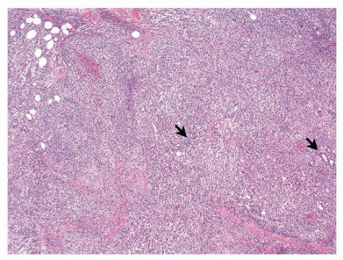
The histologic appearance is the same, regardless of patient age, and resembles YST of the gonads. A number of different histologic patterns have been described: microcystic (reticular) (Figs. 122.1, 122.2, 122.3), macrocystic, glandular-alveolar, endodermal sinus (pseudopapillary), myxomatous, hepatoid, enteric, polyvesicular vitelline, and solid10,11,12 (Fig. 122.4). There may be prominent spindling.13 The majority of YSTs show more than one histologic subtype, and the different subtypes often merge subtly from one to another. The reticular or microcystic variant is the most common histologic subtype and is characterized by a loose network of spaces and channels with small cystic spaces lined by flattened or cuboidal cells with scant cytoplasm, which may contain pale eosinophilic secretion. The nuclei vary in size but generally are small. Mitotic activity is typically brisk. A variant of the microcystic pattern is the myxomatous pattern in which the epithelial-like cells are separated by abundant myxomatous stroma. The endodermal sinus pattern has a pseudopapillary appearance and typically shows numerous Schiller-Duval bodies. These are glomeruloid structures with a central blood vessel covered by an inner rim of tumor cells, surrounded by a capsule lined by an outer (parietal) rim of tumor cells. Although characteristic of YST, Schiller-Duvall bodies are present in only 50% to 75% of YST.3 The polyvesicular vitelline pattern is composed of compact connective tissue stroma containing cysts lined by cuboidal to flat tumor cells. The solid pattern is uncommon and consists of nodular aggregates of medium-sized polygonal tumor cells with clear cytoplasm and large nuclei. Hepatoid and enteric variants are other less common forms of YST.3,14 The hepatoid pattern contains cells with abundant eosinophilic cytoplasm resembling fetal or adult liver.3,14 The enteric and endometrioid patterns show glandular features resembling the fetal human gut and endometrial glands, respectively, and have generally been reported in gonadal sites.15,16 Hyaline globules are commonly present in YST. However, hyaline droplets may also be
found in a minority of embryonal carcinomas, as well as in some other epithelial tumors. Similar to seminomas, YST can be associated with cystic changes in the thymus.
found in a minority of embryonal carcinomas, as well as in some other epithelial tumors. Similar to seminomas, YST can be associated with cystic changes in the thymus.
Stay updated, free articles. Join our Telegram channel

Full access? Get Clinical Tree


