Fig. 18.1
(a–d) Rest ECG showing a wide complex tachycardia
What are the possible types of wide QRS complex tachycardia?
Ventricular tachycardia (VT)
Supraventricular tachycardia with aberrant AV conduction
Atrial fibrillation
Atrial flutter
Focal atrial tachycardia
Atrioventricular reciprocating tachycardia (ortho- or antidromic)
Atrioventricular nodal reciprocating tachycardia
Pre-excited tachycardia (with anterograde conduction on the accessory pathway)
Atrial fibrillation
Atrial flutter
Focal atrial tachycardia
The presence of a regular rhythm excludes the hypothesis of atrial fibrillation and atrial flutter/tachycardia with variable AV conduction.
In the present ECG, there are not any visible P waves (or F wave); the P wave could be inside the terminal part of the ventricular complex or inside the T wave. So, a clear atrioventricular dissociation (suggestive for VT) is not demonstrable. Moreover, the heart rate (120 bpm) is too slow for a 2:1 atrial flutter. Even if P wave is not visible, hypothesis of a sinus tachycardia is unlikely because there were not physiological reasons for having an elevated rest heart rate.
There are not fusion or capture beats (diagnostic for VT).
There was hemodynamic stability, and therefore we performed carotid sinus massage that did not modify the tachycardia cycle. The QRS complexes in the precordial leads were not concordant: V1 positive and negative from V2 to V6 (concordance is high suggestive of ectopic ventricular beats, if negative specifically for VT). We measured the distance from the onset of QRS to the nadir of S wave in a precordial lead (we chose V2), and it is about 120 ms (when this interval is more or equal to 100 ms, you should consider VT). Finally, our ECG analysis ends with QRS morphological criteria: we are in front of a RBBB-type wide QRS tachycardia. In V1 the QRS complex morphology is Rr’ and in V6 rS. This morphological pattern is suggestive for VT. The QRS axis at −160° and the QRS duration of 200 ms are other adjuvant criteria for VT.
Owing these criteria, we postulated the diagnosis of ventricular tachycardia.
The patient, after sedation with midazolam 5 mg IV, was successfully treated with electric cardioversion (DC shock 200 J) with an immediate return to sinus rhythm (Fig. 18.2).
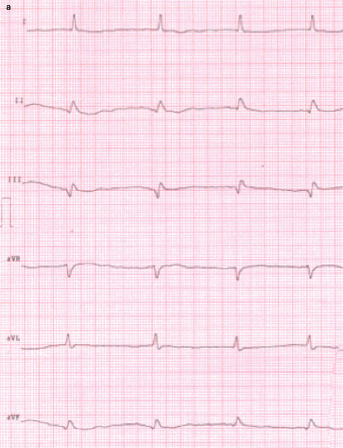
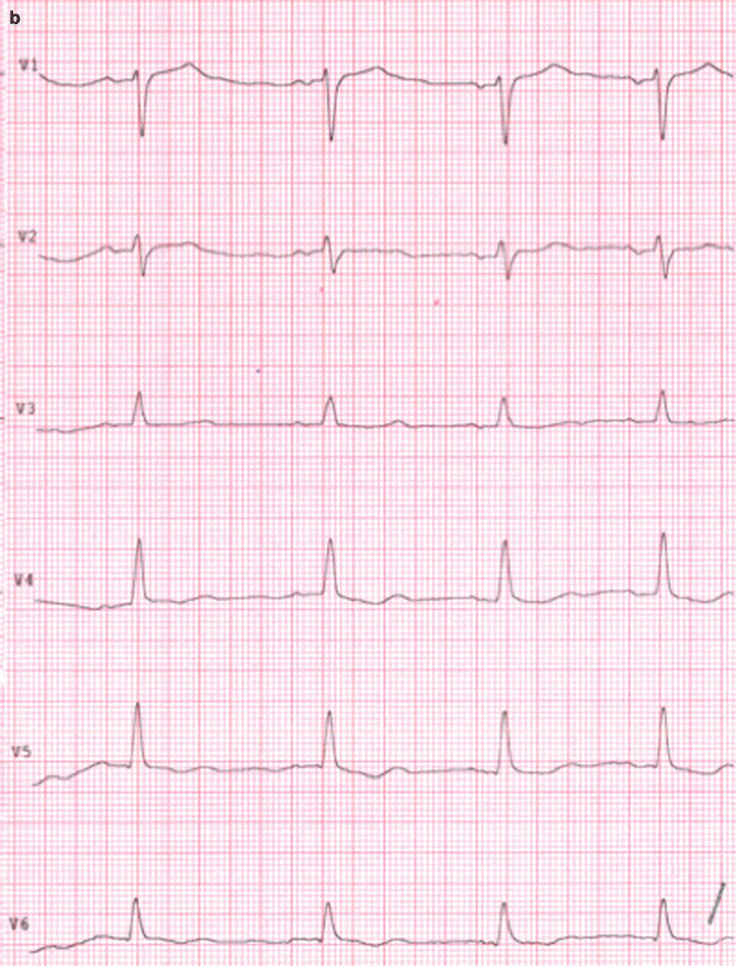
Fig. 18.2
(a, b) ECG performed after electrical cardioversion
At sinus rhythm, the QRS complex had a complete different morphology. The signs of the previous inferior MI were visible.
Admission to the Arrhythmology Department
The patient was then admitted to our cardiology and arrhythmology department to seek the cause of arrhythmia.
Electrolytec imbalance, hyper- or hypothyroidism, and coronary acute syndrome were excluded.
ECG: sinus rhythm, normal AV conduction, QRS axis +30°, Q wave in inferior leads as a previous inferior myocardial infarction, normal repolarization.
At echocardiography: mild dilatation of the left atrium. Moderate dilatation of the left ventricle (iEDV 92 ml/m2). Severe left ventricular systolic dysfunction (EF 40 %). Akinetic and thinned inferior wall of the left ventricle. Normal dimensions of the right ventricular dimension and systolic function (TAPSE 19 mm). II grade diastolic dysfunction. Moderate mitral regurgitation. Mild tricuspid regurgitation with normal pulmonary arterial pressure. No pericardial effusion.
There were not any clues attributable to accessory AV pathways or to channelopathies (e.g., Brugada syndrome or long and short QT syndrome). Also, cardiomyopathies like hypertrophic cardiomyopathy, arrhythmogenic right ventricular dysplasia, or non-compacted myocardium were excluded. Finally, the presence of an inferior wall necrosis was confirmed.
Because of the low EF unknown in previous controls, we repeated a coronary arteriography that showed occlusion of VG between RC and LMA1, occlusion of VG between aorta artery and LAD, and a good flow on LIMA to LAD.
Amiodarone therapy was started (400 mg/die for the 1st week then 200 mg/die) to prevent recurrences. During hospitalization at ECG monitor, a lot of premature ventricular beats (PVBs), couple of PVBs, non-sustained VT (nsVT), and another VT (which required cardioversion with DC shock) were recorded. All these ventricular arrhythmias had the same QRS morphology of the first VT episode.
An invasive electrophysiological study (EPS) was performed and documented:
[…] easy induction by programmed ventricular pacing of a ventricular tachycardia coming from the inferior wall of left ventricle. A catheter ablation of the site of origin of ventricular tachycardia was performed successfully with no more induction of arrhythmia after programmed ventricular pacing of the area. […]
The final diagnosis was “ventricular tachycardia starting from the inferior wall of the left ventricle due to macro-reentrant circuits secondary to a myocardial scar.”
VT did not return during hospitalization.
One month after discharge, we performed a 7-day-long ECG-Holter monitoring that did not document any VT recurrence.
18.2 Ventricular Tachycardia
Definition
VT is a tachycardia that rises from the ventricles and lasts at least 3 beats. Non-sustained VT (nsVT) is a VT that lasts less than 30 s; contrarily, a VT that lasts longer than 30 s is called sustained ventricular tachycardia (sVT).
Epidemiology and Etiology
VTs are associated with a structural heart disease, but can occur also in its absence. Ischemic heart disease is the most common cause of VT. During the acute phase of ischemia, a polymorphic VT or a ventricular fibrillation (VF) is the principal cause of sudden cardiac death (SCD).
Monomorphic VT is usually a consequence of a myocardial scar zone that became a reentrant substrate in patients with structural heart disease. The scars can derive from an old myocardial infarction but also from nonischemic cardiomyopathies, including idiopathic dilated cardiomyopathy, hypertrophic cardiomyopathy (HCM), infiltrative heart disease (e.g., amyloidosis), and arrhythmogenic right ventricular dysplasia (ARVD).
Moreover, some genetic syndromes (channelopathies) are mainly associated with polymorphic ventricular arrhythmias: Brugada syndrome, long and short QT syndromes, and catecholaminergic polymorphic ventricular tachycardia (see specific chapters).
Finally, a VT that occurs in the absence of structural heart disease and genetic conditions is referred to as idiopathic VT. This form of VTs usually originates from the right ventricular outflow tract (RVOT) and is associated with good prognosis [1].
ECG Diagnostic Criteria
In the presence of a tachyarrhythmia with wide QRS, a possible diagnosis of ventricular tachycardia (VT) has to be taken into account. The mainstay of differential diagnosis is the ECG itself, but there are other elements that may help the diagnosis.
ECG in Wide QRS Tachycardias
Wide QRS complex tachycardias are one of the most challenging questions of ECG. A regular wide QRS complex tachycardia may represent one of the following rhythms:
Aberrant atrioventricular conduction of supraventricular tachycardia (SVT)
Ventricular tachycardia (VT)
Preexisting right (RBBB) or left (LBBB) bundle branch block with SVT
Anterograde conduction over the bypass tract of atrioventricular connection in patients with Wolff-Parkinson-White (WPW) syndrome
Irregular wide QRS complex tachycardia may represent one of the following rhythms:
Atrial fibrillation with aberrant ventricular conduction< div class='tao-gold-member'>Only gold members can continue reading. Log In or Register to continue
Stay updated, free articles. Join our Telegram channel

Full access? Get Clinical Tree


