Fig. 9.1
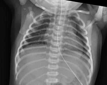
Chest x-ray showing a slightly enlarged cardiac shadow and streaky lung fields in case 1
The echocardiogram showed normal situs, atrioventricular and ventricular-arterial connections. The pulmonary veins were seen entering the left atrium. There was right to left shunting across the atrial septum. There was no forward flow across the pulmonary valve and a tortuous duct supplying confluent branch pulmonary arteries. The diagnosis of pulmonary atresia with intact ventricular septum was made and the baby was referred to the regional paediatric cardiac centre.
Case Description 2
A term baby was delivered by emergency caesarean section for a pathological cardiotocogram. There was also a history of prolonged rupture of membranes (PROM). She was born in good condition and did not require resuscitation. There was meconium present, and her APGARs were 7 at 1 min and 9 at 5 min.
The paediatrician was asked to review her because she failed pulse oximetry screening on the postnatal ward. Her saturations were 95 % pre-ductal and 92 % post-ductal. She was brought to the neonatal unit where she was started on 6 L oxygen via optiflow. Her oxygen saturations improved, with pre-ductal reaching 100 % and post-ductal reaching 100 %. Again, given the history of meconium-stained liquor at delivery, she was thought to have meconium aspiration syndrome. She went on to have an infection screen and was started on intravenous first-line antibiotics. Initial capillary blood gas was pH of 7.33, pCO2 of 5.8 kPa and base excess of 2.6.
As she was requiring 100 % oxygen, the decision was made to intubate and ventilate her. However, on consultant review she had no signs of respiratory distress and was settled in 6 L of 100 % oxygen via optiflow. Repeat blood gas showed a pH of 7.44, pCO2 of 4.56 kPa and BE −0.2. Chest x-ray on admission is shown in Fig. 9.2. Given that this was normal, she was suspected of having early PPHN secondary to sepsis, as there was a history of PROM. Intubation was avoided and an echocardiogram showed a structurally normal heart with evidence of mild PPHN. There was predominantly right to left shunt across the PDA and the patent foramen ovale as shown on Fig. 9.3.
 < div class='tao-gold-member'>


Only gold members can continue reading. Log In or Register to continue
Stay updated, free articles. Join our Telegram channel
Full access? Get Clinical Tree
