Weight Loss, Fatigue, Increasing Dyspnea, and Leg Edema
A 54-year-old man has suffered from weight loss and fatigue for 3 to 6 months and increasing dyspnea for 6 weeks.
Leg edema occurred over the last 4 weeks, and the patient was referred for an echocardiographic workup.
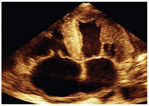
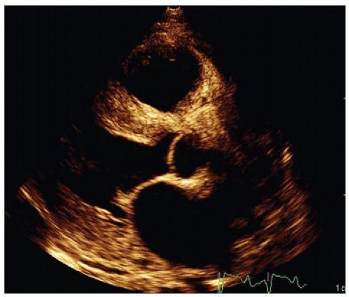
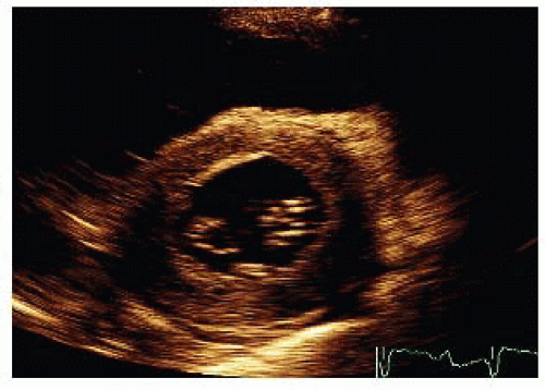
The transthoracic echocardiogram images in Videos 35-1 to 35-3 and Figures 35-1, 35-2 and 35-3 were acquired.
QUESTION 1. Which is the most likely diagnosis according to these echocardiogram findings and the medical history?
A. Hypertrophic cardiomyopathy
B. Hypertension
C. Cardiac amyloidosis
D. Left ventricular noncompaction
View Answer


ANSWER 1: C. These echocardiographic images show the typical appearance of cardiac amyloidosis in an advanced stage of the disease.
The typical features—a concentric left and right ventricular thickening, a normal left ventricular cavity size, dilated right and left atria, and a tiny pericardial effusion—can be observed in this patient.1 The left ventricular systolic function is usually preserved up to a late stage of the disease. Diastolic dysfunction normally occurs early in cardiac amyloidosis and can be assessed by using standard and Doppler echocardiography.
A myocardial texture appearance with “granular sparkling” allows for the diagnosis of a cardiac amyloidosis with a sensitivity of 87% and specificity of 81%.2
Stay updated, free articles. Join our Telegram channel

Full access? Get Clinical Tree


