Fig. 28.1
The Amplatzer Muscular VSD Occluder, AGA Medical Corporation, St. Jude, MN. A device size/waist diameter (mm), B disc diameter (mm), C waist length (mm)
28.3.2 Device for PMVSD
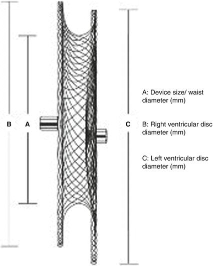
The Amplatzer membranous ventricular septal defect occluder has two discs of unequal size (Amplatzer Membranous VSD Occluder, AGA Medical Corporation, St. Jude, MN). The aortic rim of the asymmetric left ventricular disc exceeds the dimensions of the connecting waist by only 0.5 mm, so as to avoid impingement on the aortic valve, whereas the apical end is 5.5 mm larger than the waist. This apical end of the left ventricular disc contains a platinum marker to facilitate correct orientation during implantation. The right ventricular disc is symmetrical, and it exceeds the diameter of the connecting waist by 2 mm throughout its circumference (Fig. 28.2). The device is available in sizes from 4 to 18 mm and requires delivery sheaths from 7 to 9 French.


Fig. 28.2
The Amplatzer Membranous VSD Occluder, AGA Medical Corporation, St. Jude, MN. A device size/waist diameter (mm), B right ventricular disc diameter (mm), C left ventricular disc diameter (mm)
A MVSD Amplatzer device II is also available. It was redesigned to prevent conduction abnormalities with a 75 % reduction in radial force, 45 % reduction in clamping force and 10 % increase in stability. The left disc of this new device has an elliptical and concave shape that adapts to the LV outflow tract. It is available in two configurations:
1.
Eccentric with a 1-mm superior rim and a 3-mm inferior rim
2.
Concentric with a 3-mm superior and inferior rims
The waist length is increased from 1.5 to 3 mm, and the waist diameter ranges from 4 to 10 mm in 1-mm increments plus 12 and 14 mm. Polyester patches are sewn into the discs.
The delivery system consists of a delivery cable and a pusher catheter having a sharp curvature of 180° inferiorly. This allows correct orientation of the left ventricular disc during implantation. It has a flattened part of the socket that matches the flat portion of the microscrew, in order to force larger part of the left ventricular disc to be oriented downwards so that it points to the left ventricular apex.
28.4 Procedure
28.4.1 Preparation
General anaesthesia and orotracheal intubation.
Biplane catheterization laboratory preferred.
Patient position with arms lifted up, behind patient’s neck (attention to brachial plexus overstretching).
Patient is fully monitored including an arterial line for continuous arterial pressure monitoring, two peripheral venous lines or a central venous line and vesical catheter for diuresis evaluation.
A transoesophageal echocardiography 2D (2D-TEE) or 3D (3D-TEE) must be used to monitor the procedure.
Heparinization with IV administration of 100 UI/kg heparin. Check hourly the activated clotting time >250 s. In case administer heparin intravenously during the procedure. Usually it is not needed.
Antibiotics IV: usually a cephalosporin.
The procedure has to be considered as a surgical intervention. Therefore, special care has to be paid to strict asepsis. Special attention has to be given to operators’ scrubbing and patient’s preparation (including careful depilation). The personnel involved has to wear masks and hats.
28.5 Access Site
A femoral vein (FV) access is used to approach the closure of a PMVSD, and an internal jugular vein (IJV) can be used for MVSD closure.
An arteriovenous circuit must be created (internal jugular vein-femoral artery for MVSD and femoral vein-femoral artery for PMVSD closure).
Both sides for vascular femoral access are prepared.
28.6 Catheterization and Haemodynamic Evaluation for MVSD Closure
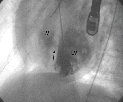
Left ventricular angiographies are obtained in axial projections for best evaluation of VSD size and position, in addition to TEE views. Left ventriculography in the hepatoclavicular projection (35° left anterior oblique/35° cranial) is performed to imaging mid-muscular, apical posterior defects. Anterior defects are better seen in 60° left anterior oblique/20° cranial (Fig. 28.3).


Fig. 28.3
Left ventricular angiogram: LV left ventricle, RV right ventricle, Arrow mid muscular VSD
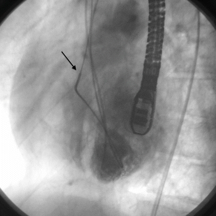
The VSD is crossed from the left side by using a right Judkins catheter and a soft Glidewire (0.035″, J Tip, Terumo); the wire is advanced to the pulmonary artery or to the SVC/IVC, where it is snared with a GooseNeck Snare (Microvena Corporation, 20–25 mm in adults, 10–15 mm in children) and exteriorized out of the right internal jugular vein or femoral vein establishing an arteriovenous circuit (Fig. 28.4).


Fig. 28.4
Left ventricular angiogram: the arrow shows the arteriovenous circuit (femoral artery-internal jugular vein)
Over the circuit, an appropriate size delivery sheath is advanced from the vein all the way until the tip of the sheath is in the ascending aorta. The dilator is withdrawn, and the sheath is pulled back in the left ventricle.
When the tip of the sheath is placed in the mid cavity of the left ventricle, the dilator and the wire are gently removed; a left ventriculogram is usually repeated, to confirm the position of the long sheath and also to obtain additional information on the position and the size of the VSD.
According to both angiographic and echocardiographic information, a muscular VSD occluder 1–2 mm larger than the maximum size of the defect is chosen.
The device is attached to the delivery cable, loaded into the plastic loader, introduced and advanced into the sheath.
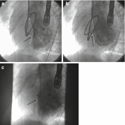
The left disc is deployed in the left ventricular cavity, making sure it is not impinged in mitral valve apparatus, then the entire system is withdrawn towards the septum (Fig. 28.5a), and the central waist and the proximal disc are deployed; a test angiogram is done to verify the correct position of the device (Fig. 28.5b).


Fig. 28.5
Left ventricular angiogram: (a) The system is withdrawn towards the septum (arrow); (b) the central waist and the proximal disc are deployed (arrow); (c) a test angiogram is done to verify the correct position of the device (arrow). LV left ventricle
< div class='tao-gold-member'>
Only gold members can continue reading. Log In or Register to continue
Stay updated, free articles. Join our Telegram channel

Full access? Get Clinical Tree


