Venous Thromboembolism


Venous Thromboembolism


Cynthia Wu

Mark A. Crowther


Venous thromboembolism (VTE) comprises of both deep vein thrombosis (DVT) and pulmonary embolism (PE). VTE is associated with significant morbidity and mortality. Fully 10% of symptomatic PE is fatal within the first hour of presentation. Because there are extremely effective therapies to both prevent and treat VTE, it is important to be able to recognize the signs and symptoms of DVT and PE, to be aware of the clinical situations in which a high index of suspicion is warranted, to have a simple and accurate diagnostic algorithm, and to understand the various options for treatment.

VTE is age dependent, varying from an annual incidence of 0.03% in subjects under 50 years of age to 0.4% in those over 50 years (incidence of 1 per 10,000 at age 25 to 1 per 1,000 at age 65). Most studies have reported an equal incidence for both sexes. The majority of DVT is diagnosed in the lower extremities, often starting in the calf veins. Proximal DVT has a much stronger association with PE, and it is estimated that 50% of patients with untreated proximal DVT or PE develop recurrent VTE within 3 months. Isolated calf vein thrombi rarely cause clinically significant complications such as PE, and the majority of calf vein thrombi (when detected using screening techniques) resolve spontaneously even in the absence of anticoagulant therapy. About 25% of untreated calf DVT extends proximally, usually within a week of presentation. About half of patients with symptomatic proximal DVT have evidence of PE on imaging studies even without clinically evident respiratory symptoms, and about 70% of patients with symptomatic PE have DVT (2/3 are proximal).


CLINICAL FEATURES OF VENOUS THROMBOEMBOLISM

The clinical findings in DVT and PE are both insensitive and nonspecific. Physical examination must be used in conjunction with a thorough history to rule out alternative diagnoses. The diagnosis requires an evaluation of VTE risk factors and the appropriate use of diagnostic tools. Occasionally, events are entirely asymptomatic and found incidentally on imaging studies done for other reasons. Common clinical presentations for DVT and PE include the following:


Deep Vein Thrombosis


  • leg swelling +/- pitting edema


    • particularly if unilateral or involving the whole leg


    • objective measurement of circumference best taken 10 cm below the tibial tuberosity and compared to the unaffected leg


  • leg pain


    • distribution of the deep veins—within the calf and medial thigh


    • localized tenderness, particularly over a palpable cord or visible group of vessels, may be more in keeping with superficial phlebitis


  • redness and warmth


  • pallor or cyanosis (rarely)


    • may be associated with massive occlusive iliofemoral DVT


  • Homan’s sign (calf pain with forced dorsiflexion of the foot) is neither sensitive nor specific for DVT

Other diseases may present with the same symptoms. Some of these include leg trauma, cellulitis, ruptured Baker’s cyst, venous insufficiency, external compression of local vessels or lymphatics, and superficial phlebitis. Patients with postphlebitic syndrome from previous DVT may also have chronic leg swelling and pain.


Pulmonary Embolism


  • chest pain, particularly pleuritic, sudden in onset and persistent


  • sudden onset of shortness of breath


  • preceding symptoms of DVT


  • tachycardia, cyanosis, signs of right-sided heart failure


  • hemoptysis (rare)


    • secondary to pulmonary infarction


  • syncope/shock (rare)


    • secondary to obstruction of right-sided cardiac output

Other causes for dyspnea and chest pain should be promptly ruled out (e.g., myocardial infarction, congestive heart failure, pneumonia) as many of these also carry significant morbidity and mortality in the absence of appropriate treatment. Patients with previous PE who develop pulmonary hypertension that does not resolve after the acute event may also suffer from chronic chest pain and shortness of breath.


RISK FACTORS FOR VENOUS THROMBOEMBOLISM

When a patient presents with symptoms suggestive of DVT or PE, it is often useful to determine the background risk for developing VTE as this not only guides further diagnostic testing but also may affect the aggressiveness and the duration of any subsequent treatment. Approximately 25 to 50% of first events are idiopathic in nature with no obvious provoking incident. Despite this, there exists a wide spectrum of known factors that increase the risk of VTE. In 1856, German physician Rudolf Virchow devised a simple framework for categorizing these factors. This is commonly known as Virchow’s triad and consists of three pathophysiological mechanisms: stasis, endothelial injury, and hypercoagulability. Thrombotic risk can alternatively be seen as either inherited or acquired. Common risk factors for VTE can be summarized using these approaches (Table 21.1). It is also important to determine whether or not the contributing risk factors are transient or permanent and if any can be attenuated with other
VTE-unrelated interventions. Controlling or eradicating underlying risk factors can decrease the chances of treatment failure and thrombotic recurrence and can possibly shorten the duration of anticoagulation.


TABLE 21.1 RISK FACTORS FOR VTE


Venous Stasis


Prolonged immobility


Hospitalization, stroke, paralysis


Fracture with cast/non-weight-bearing status


External compression of local vasculature and lymphatic channels


Endothelial Injury


Major trauma


Major surgery


Intravenous catheters (central venous access)


Hypercoagulability


Acquired


Cancer and chemotherapy


Pregnancy


Hormone therapy (oral contraceptive, hormone replacement)


Antiphospholipid antibody syndrome


Heparin-induced thrombocytopenia


Inherited


Factor V Leiden


Prothrombin 20210 gene mutation


Protein C deficiency


Protein S deficiency


Antithrombin deficiency


Special Patient Groups


Hospitalized Medical Patients

Most hospitalized medical patients have at least one risk factor for VTE (usually immobility), and most have multiple risks relating to their reason for admission and other comorbid diseases. Without prophylaxis, venographically detected DVT can occur in 10 to 20% of such patients, and when symptomatic, VTE may contribute significantly to in-hospital morbidity, mortality, and length of stay. Preventative measures should be considered in all patients without contraindications to anticoagulation, and a high index of suspicion should be maintained for VTE in these patients.


Hospitalized Surgical Patients

Surgical patients generally have a higher risk of VTE than medical patients, especially trauma patients and those requiring orthopedic procedures such
as hip or knee replacements. In such patients, the rate of venographically detected VTE may be as high as 40 to 60% without prophylaxis. After hemostasis is achieved postoperatively, DVT prophylaxis should be initiated in all patients with an important risk of DVT. In some situations, prophylaxis may extend beyond discharge. A high index of suspicion must be maintained if any symptoms of VTE occur.


Cancer Patients

Cancer and its treatment are independent risk factors for VTE and increase the risk fourfold to sixfold. About 15 to 20% of patients with acute VTE concurrently have a diagnosis of cancer, and 10 % of patients with idiopathic VTE are diagnosed with cancer within the first year after their thrombotic event. VTE in cancer patients is associated with a more aggressive course, more frequent treatment failures, increased bleeding risk, and shortened life expectancy. Diagnosis may be delayed due to multiple competing alternative explanations for the presenting symptoms. As the risk of VTE is high in these patients, VTE is often considered in the differential diagnosis.


Antiphospholipid Antibody Syndrome

Antiphospholipid antibody syndrome (APLAS) can be a powerful stimulus for both large and small vessel arterial and venous thromboses. DVT is one of the common presenting manifestations of APLAS. The diagnostic criteria for APLAS are provided below and require one clinical criterion and one laboratory criterion. Treatment may also be complicated in patients with concurrent moderate or severe thrombocytopenia.


Clinical Criteria


  • Vascular thrombosis (venous/arterial/small vessel) in any tissue/organ. Superficial venous thrombosis is not included in clinical criteria.


  • Pregnancy morbidity


Laboratory Criteria


  • Lupus anticoagulant present in plasma/serum >2 occasions at least 12-weeks apart


  • Anticardiolipin antibodies (IgG and IgM) in plasma/serum > 2 occasions at least 12-weeks apart


  • Anti-β2 glycoprotein 1 antibodies (IgG and IgM) in plasma/serum > 2 occasions at least 12-weeks apart


Hereditary Thrombophilia

The list of inherited conditions that increase the risk of thrombosis continues to grow. More common conditions such as Factor V Leiden are usually associated with an excess of procoagulant proteins and are generally weaker risk factors for the development of VTE. Rarer conditions such as protein C, protein S, and antithrombin III (AT III) deficiencies are usually associated with deficiencies of anticoagulant proteins and are generally stronger risk factors for the development of VTE. While most of these conditions have been shown to increase the risk of a first venous
thrombotic event, not all are strongly linked to an increased risk of recurrent events. Thus, caution must be used while interpreting the results of these tests, and widespread screening in all patients with VTE is not recommended.

Testing for thrombophilia may be considered in the following situations:


  • First presentation at a young age


  • Strong family history of VTE


  • Recurrent idiopathic VTE and VTE in unusual sites (portal, splenic, mesenteric, or cerebral vein thromboses)


  • Extensive VTE

It is also important to note that anticoagulant use can interfere with accurate interpretation of some thrombophilia testing (e.g., use of warfarin will decrease the normal levels of protein C and protein S even without a hereditary deficiency), and thus timing of testing should also be considered carefully. Other medical conditions such as nephrotic syndrome, liver failure, and pregnancy may also alter the normal levels of procoagulant and anticoagulant proteins even in the absence of hereditary deficiencies. In such patients, thrombophilia testing should either not be done or be done and the results interpreted with extreme caution.


DIAGNOSIS OF VENOUS THROMBOEMBOLISM

The accurate diagnosis of VTE relies on the appropriate use of a combination of tools starting from clinical assessment (focused history and physical examination) to determine the “pretest probability” of DVT or PE and judicious use of laboratory investigations and imaging modalities (Figs. 21.1 and 21.2).


Clinical Assessment

The most commonly used clinical prediction models for VTE are the Well’s Criteria for DVT and PE (Tables 21.2 and 21.3). Using a few simple and objective features from the patient’s history and physical exam, the clinician can stratify patients as low, moderate, or high probability for VTE. The only room for subjectivity lies in the score for “alternative diagnosis.” If no clearly defined alternate diagnosis can be given, patients should be scored accordingly. These clinical prediction models have been incorporated into many diagnostic algorithms to aid in deciding what, if any, further testing should be done (Figs. 21.1 and 21.2).


Laboratory Investigations

The only currently available and widely used laboratory test for the diagnosis of VTE is the d-dimer assay. All d-dimer assays rely on the use of monoclonal antibodies to d-dimer molecules. The d-dimer, two covalently bound D-domains of cross-linked fibrin monomers, is a product of fibrin degradation. They can be found circulating at low levels under physiologic conditions as well as at elevated levels in a number of disease states associated with fibrin formation and fibrinolysis. Thus, the presence of d-dimer
is not specific for venous thrombosis with the specificity reduced in patients with cancer, pregnant women, and hospitalized and elderly patients. Techniques that detect d-dimer include enzyme-linked immunosorbent assays, latex agglutination and immunoturbidimetric tests, and whole blood agglutination methods. Specific assays that have been validated for use in VTE diagnosis include (i) SimpliRED d-dimer (Agen Biochemical Ltd., Brisbane, Australia), (ii) Vidas d-dimer (bioMérieux, France), (iii) MDA d-dimer (bioMérieux, Durham, NC), and (iv) Tiniquant d-dimer (Roche Diagnostics, The Netherlands). Results of each test cannot be generalized to other methods of d-dimer quantification due to the wide variety of assay techniques. Generally, though, all are characterized by an intermediate to high sensitivity coupled with an intermediate to low specificity. This allows a negative d-dimer, in combination with a low clinical pretest probability, to effectively rule out acute VTE (less than 1% chance that a patient has VTE). Positive d-dimer results, however, do not equate to a diagnosis of VTE, and further testing is required.


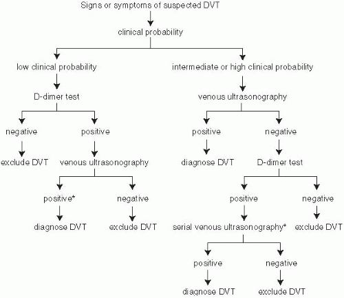
FIGURE 21-1. Diagnostic algorithm for deep vein thrombosis. (From Hirsh J, Lee AYY. How we diagnose and treat deep vein thrombosis. Blood 2002;99:3102-3110.)


Biomarkers in Risk Stratification of Venous Thromboembolism

Use of d-dimer testing to stratify patients for their risk of recurrent events is still being investigated, although preliminary results in this area are
promising. d-dimer testing may help to make decisions on whether to stop or discontinue anticoagulation therapy. Troponin and BNP measurements may help to stratify patients with PE for their risk, with several studies
showing an association between elevation and short-term risk for dying and recurrent PE. Conversely, the absence of elevation in these parameters is a favorable prognostic factor, with limited studies suggesting an almost 100% negative predictive value for complications if plasma BNP levels are normal. The use of troponin with RV functional abnormalities may also be helpful with the absence of right ventricular dysfunction and a normal troponin level identifying patients who are eligible for early discharge or even outpatient treatment.


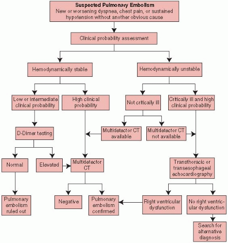
FIGURE 21-2. Diagnostic algorithm for PE. The initial assessment of the clinical probability of PE is based on either clinical judgment or clinical decision rules (e.g., Well’s Criteria). Patients are considered to be hemodynamically unstable if they are in shock or have a systolic blood pressure of less than 90 mm Hg or a drop in pressure of more than 40 mm Hg for more than 15 minutes (in the absence of new-onset arrhythmia, hypovolemia, and sepsis). In cases in which multidetector CT is not available or in patients with renal failure or allergy to contrast dye, the use of ventilation-perfusion scanning is an alternative. In patients with a high clinical probability and an elevated d-dimer level but with negative findings on multidetector CT, venous ultrasonography should be considered. Among critically ill patients with right ventricular dysfunction, thrombolysis is an option; multidetector CT should be performed when the patient’s condition has been stabilized if doubts remain about clinical management. In patients who are candidates for percutaneous embolectomy, conventional pulmonary angiography can be performed to confirm the diagnosis of PE immediately before the procedure, after the finding of right ventricular dysfunction. (From Agnelli G, Beccatini C. Current concepts. Acute pulmonary embolism. N Engl J Med 2010;363:266-274, with permission.)


TABLE 21.2 CLINICAL PREDICTION RULE FOR PREDICTING DVT


Clinical Feature


Score


Active cancer (treatment ongoing or within previous 6 mo or palliative)


1


Paralysis, paresis, or recent plaster immobilization of the lower extremities


1


Recently bedridden for more than 3 d or major surgery, within 4 wk


1


Localized tenderness along the distribution of the deep vein system


1


Entire leg swollen


1


Calf swelling by >3 cm when compared with the asymptomatic leg (measured 10 cm below tibial tuberosity)


1


Pitting edema (greater in the symptomatic leg)


1


Collateral superficial veins (nonvaricose)


1


Alternative diagnosis as likely or greater than that of DVT


-2


Total:


In patients with symptoms in both legs, the more symptomatic leg is used. Pretest probability calculated as the total score: high ≥ 3; moderate 1 or 2; low ≤ 0


Adapted from Wells PS, Anderson DR, Bormanis J, et al. Value of assessment of pretest probability of deep-vein thrombosis in clinical management. Lancet 1997;350:1795-1798.)


TABLE 21.3 CLINICAL PREDICTION RULE FOR PREDICTING PE


Variable


Points


Immobilization or surgery in the previous 4 wk


1.5


Malignancy (treatment ongoing or within previous 6 mo or palliative)


1.0


Previous DVT/PE


1.5


Hemoptysis


1.0


Heart rate > 100 beats/min


1.5


Clinical signs and symptoms of DVT (leg swelling and pain with palpation of deep veins)


3.0


An alternative diagnosis is less likely than PE


3.0


Total:


Low probability < 2.0; moderate probability 2.0-6.0; high probability ≥ 6.0.


(Adapted from Wells PS, Anderson DR, Rodger M, et al. Derivation of a simple clinical model to categorize patients probability of pulmonary embolism: increasing the models utility with the SimpliRED D-dimer. Thromb Haemost 2000;83:416-420.)


Imaging Modalities


Reference Standard Tests

In order to standardize the approach in evaluating diagnostic tools for VTE, it is often useful to think about the probability of developing VTE if the test was negative. For a test to be helpful, the diagnostic modality should have excellent negative predictive value with a ≤1 % chance of subsequently developing symptomatic VTE over a 3-month observation period if testing is negative. Sensitivity and specificity of all other imaging modalities are measured against these standards.


Contrast Venography for Deep Vein Thrombosis

The high sensitivity and specificity of combining other diagnostic tools have made use of venography less frequent for proximal DVT. It remains a fairly reliable test to diagnose calf DVT, but this is offset by its invasiveness and exposure of patients to the adverse effects of IV contrast dye and radiation. Venography is reserved for patients with otherwise nondiagnostic testing where the test results may change the management.

Only gold members can continue reading. Log In or Register to continue

Stay updated, free articles. Join our Telegram channel

Jun 12, 2016 | Posted by in CARDIOLOGY | Comments Off on Venous Thromboembolism

Full access? Get Clinical Tree

Get Clinical Tree app for offline access