Vasculogenic Erectile Dysfunction


Vasculogenic Erectile Dysfunction


Gregory Lowe

Robert Bahnson


Erectile dysfunction (ED) is defined as the persistent inability to achieve and maintain penile erection sufficient for sexual intercourse. The incidence of ED increases with age. Approximately 1 to 10% of men aged 40 to 59 years have ED compared to 20 to 30% of men greater than 60 years of age. This prevalence has been confirmed in multiple international studies. ED is now well recognized as an early marker for systemic vascular and coronary disease and indeed shares many of the same risk factors (Table 24.1). As research emerges correlating ED with the future risk of coronary artery disease and systemic vascular disease, these patients represent a group for preventative care. With the introduction of oral treatment for ED, public awareness and the number of men seeking treatment have risen. The initiation of medical therapy for ED is increasingly being administered in the primary care physician’s office, with those patients failing oral therapy obtaining specialist referral.


CLINICAL FEATURES


Physiology and Pathophysiology of an Erection


Vascular Supply and Innervation of the Penis

An understanding of the anatomy and physiology of normal penile erection is crucial to appropriate identification of likely etiologies and planning treatment strategies. Attainment of an erection results from multiple influences converging on the penile smooth muscle. An erection requires adequate blood flow to the paired corpora cavernosae surrounding the penile urethra and the corpus spongiosum at the glans penis (Fig. 24.1).

The central nervous system plays an important role in erectile function. The hypothalamus (medial preoptic area) is the integration point for the central control of erection. This area receives rich sensory impulses from a multitude of brain areas. Auditory stimuli, visual stimuli, and fantasy-initiated impulses project to this area and then travel down the spinal cord to peripheral nerves that innervate the penile vasculature initiating an erection. The penis is innervated by the sympathetic (T11-L2), parasympathetic (S2-S4), and somatic (S2-S4) nervous systems. Sympathetic input is antierectile, whereas parasympathetic and somatic inputs are proerectile. Both sympathetic and parasympathetic fibers reach the inferior hypogastric plexus in the pelvis, where the autonomic input to the penis is integrated and penile innervation via the cavernous nerves originates. The lesser cavernous nerves travel along the penis to supply the penile urethra and erectile tissue of the corpus spongiosum, while the greater cavernous nerves innervate the helicine arteries in the erectile
tissue. The somatic component, the pudendal nerve, through the dorsal nerve branches is responsible for penile sensation and the contraction and relaxation of the extracorporeal striated muscles (bulbocavernous and ischiocavernous).


TABLE 24.1 RISK FACTORS ASSOCIATED WITH ED


Hypertension


Hyperlipidemia


Coronary artery or peripheral arterial disease


Diabetes


Metabolic syndrome


Hypogonadism


Endocrine disorders


Smoking


Depression


Alcohol and drug abuse


Peyronie’s disease


Trauma or surgery to the pelvis or spine


Vascular surgery


Erection Phase

In response to sexual stimulation, parasympathetic neural activity predominates, resulting in increased blood flow into the cavernous sinuses and achievement of erection due to smooth muscle relaxation. The main neurotransmitter mediating smooth muscle relaxation is nitric oxide, which is released from the endothelium of the corpus cavernosa during nonadrenergic, noncholinergic neurotransmission. Nitric oxide, in turn, activates soluble guanylyl cyclase, which increases intracellular concentrations of cyclic guanosine monophosphate (cGMP). cGMP, in turn, results in the activation of cGMP-dependent protein kinase, which, in turn, phosphorylates specific proteins and ion channels. The consequence is a decrease in intracellular calcium concentration and relaxation of the smooth muscle. The rapid filling of the penile sinusoids results in expansion of the sinuses with resultant compression of the subtunical venular plexuses, resulting in near total occlusion of venous outflow. This results in trapping of blood within the corpus cavernosum raising the penis from a dependent position to an erect position. Resultant intracavernosal pressures may exceed 90 mm Hg during the full phase of penile erection. During the course of sexual intercourse or masturbation, the bulbocavernous reflex results in forcible compression of the base of the blood-filled corpora cavernosa, resulting in the penis becoming harder with intracavernous pressures exceeding several 100 mm Hg. During this maintenance phase of penile erection, both blood entering and blood leaving the penis temporarily seize.


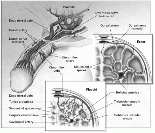
FIGURE 24-1. Anatomy and mechanism of penile erection. The mechanisms of erection and flaccidity are shown in the upper and lower inserts, respectively. During erection, relaxation of the trabecular smooth muscle and dilatation of the arterioles result in an increase in blood flow, which expands the sinusoidal spaces to lengthen and enlarge the penis. The expansion of the sinusoids compresses the subtunical venular plexus against the tunica albuginea and simultaneously compresses the emissary veins, reducing the outflow of blood to a minimum. (From Lue TF. Erectile dysfunction. N Engl J Med 2000;342:1802-1813, with permission.)


Detumescence Phase

This results from the sudden suppression of neurotransmitter release, breakdown of second messenger products, or discharge of sympathetic stimuli during ejaculation. Constriction of the trabecular smooth muscles reopens the venous channels resulting in expulsion of the trapped blood and return of a flaccid state. ED may result in abnormalities in one or several of these steps involved in the initiation or maintenance of an erection.


Future Cardiovascular Risk in Patients with Erectile Dysfunction and Cardiovascular Risk of Sexual Activity

The Second Princeton Consensus Panel is a practice guideline for the management of ED in patients with significant cardiac risk, which stratifies patients according to the presence or severity of conventional cardiovascular risk factors [low risk (<10%), intermediate (10 to 20%), and high risk
(>20%)]. A careful assessment of subclinical and/or asymptomatic cardiovascular disease should be considered, especially in patients with ED and an estimated cardiovascular risk greater than 10%.

Several studies have looked at the risk of cardiovascular events with sexual activity. After correcting for chance, only 0.9% of cases presenting with acute MI are currently felt to relate to antecedent sexual activity. This risk is exceedingly low in patients able to exert at least six metabolic equivalents of oxygen consumption (METS). Man-on-top intercourse leads to the highest heart rate with a mean peak heart rate of 127 beats/min. Overall vigorous sexual activity is estimated to equate to about 5 to 6 METS, with a mean 3.3 METS at orgasm for men-on-top intercourse. Masturbation and woman-on-top position are associated with lower metabolic demand.


Differential Diagnosis

The differential diagnosis of ED includes a variety of etiologies (Table 24.2). Aside from organic psychiatric causes of ED, a number of well-recognized stressors can trigger performance anxiety including anger, alienation, and depression. Mental illness and drugs used to treat them can commonly cause ED. Commonly prevalent risk factors that are implicated in atherosclerosis such as smoking, high cholesterol, and diabetes mellitus can all cause ED. Systemic disorders such as diabetes, chronic renal failure, and even aging can commonly contribute to ED via a combination of factors including neuronal and vascular dysfunctions.


Organic Causes of Erectile Dysfunction

Neurologic. Damage to the CNS from cerebrovascular accidents and degenerative processes such as Alzheimer’s or Parkinson’s disease may all result in ED. Similarly, damage to the peripheral nervous system as may ensue from spinal cord injury, diabetic neuropathy, and vascular or prostate surgery are common causes of ED.

Endocrine. Testosterone deficiency causes both decreased libido and an inability to maintain an erection secondary to low levels of nitric oxide synthase. In addition, hyperprolactinemia, hyperthyroidism, and hypothyroidism can all cause ED. In these settings, normalizing hormonal levels often results in return of normal male sexual function.

Vasculogenic. Anatomic defects in the arterial and venous systems may cause ED. Arterial stenosis and vasodilator dysfunction secondary to atherosclerosis can prevent adequate blood flow to the penis for an erection and are the most common causes of ED. Arterial stenosis involving the aortoiliac blood vessels resulting in claudication in one or both legs, diminution in femoral pulses, and ED is referred to as Leriche’s syndrome. Venous dysfunction causing failure of veins to close during an erection may prevent the maintenance of an erection. Causes of abnormal venous drainage include Peyronie’s disease, aging, and diabetes mellitus.


TABLE 24.2 RISK FACTORS ASSOCIATED WITH ED DIFFERENTIAL DIAGNOSIS OF ED


Common Disorders


Pathophysiology


Psychogenic


Depression


Decreased libido


Anxiety disorder Schizophrenia Relationship conflict Stress


Treatment side effects


Organic


Neurologic


Cerebrovascular accident


Failure to initiate or transmit


Alzheimer’s or Parkinson’s disease Spinal cord injury Diabetic neuropathy Radical pelvic surgery


nerve impulses


Hormonal


Hypogonadism


Decreased libido


Thyroid disease Hyperprolactinemia


Endothelial dysfunction


Vascular


Atherosclerosis


Endothelial dysfunction


Peyronie’s disease


Decreased arterial blood flow


Trauma


Impaired veno-occlusion


Drug induced


Antidepressants


Central suppression


Antihypertensives Diuretics Alcohol and cigarette abuse


Decreased libido


Multifactorial


Systemic disease


Diabetes mellitus


Vascular dysfunction


Chronic renal failure Coronary artery disease


Neural dysfunction


Mixed psychogenic and organic


Mixture of above


Multifactorial


(Adapted from Lue TF. Erectile dysfunction. N Engl J Med 2000;342:1802-1813.)


Medications. Table 24.3 lists common medications that should be considered during the evaluation of ED.


Erectile Dysfunction and Vascular Risk Factors

ED and lower-extremity arterial disease share many of the same risk factors. These include diabetes mellitus, metabolic syndrome, smoking, hypertension, dyslipidemia, obesity, and age, all of which are well-known

Only gold members can continue reading. Log In or Register to continue

Stay updated, free articles. Join our Telegram channel

Jun 12, 2016 | Posted by in CARDIOLOGY | Comments Off on Vasculogenic Erectile Dysfunction

Full access? Get Clinical Tree

Get Clinical Tree app for offline access