(1)
Department of Neuroscience, University of Turin Ospedale Molinette, Turin, Italy
Abstract
In this chapter, the vascular territories of the brain as viewed in the axial planes on CT or MRI are presented.
In this chapter, the vascular territories of the brain as viewed in the axial planes on CT or MRI are presented.
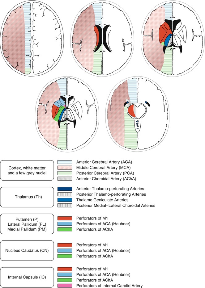
The cerebral hemispheres are vascularized by the anterior cerebral (ACA), middle cerebral (MCA), posterior cerebral (PCA), and anterior choroidal (AchA) arteries. From the proximal segments of these arteries (A1, M1, P1) and cisternal segment of the choroidal artery, small branches (deep perforators) arise that enter the brain parenchyma at the level of the anterior and posterior perforated substance supplying basal ganglia, thalamus, and internal capsule. Other perforators arise from the anterior communicating (AComA) and posterior communicating (PComA) arteries, which belong to the circle of Willis, and from the distal segment of the internal carotid artery. The perforators are end-arteries with no possible collateral circulation. From the more distal branches of ACA, MCA, PCA, and AchA, cortical arteries that supply the cortex and arteries that enter the white matter (medullary arteries) arise. Like the deep perforators, the medullary arteries are also end-arteries. On the contrary, there are potential pial (leptomeningeal) anastomoses among the cortical branches of all the main trunks (Fig. 8.1).