Vascular Applications in Point of Care Ultrasound
R. Eugene Zierler
The use of ultrasound for medical diagnosis began in the late 1940s and early 1950s based on experience with SONAR (sound navigation and ranging) and metal flaw detection devices. These military and industrial applications of ultrasound paved the way for research on ultrasound as a noninvasive tool for characterizing biologic tissues. However, commercial ultrasound equipment did not become available until the 1960s, and those early devices were limited to A-mode (amplitude mode) displays that produced a one-dimensional tissue image and continuous-wave (CW) Doppler for detection of blood flow.1,2 Two-dimensional gray-scale imaging was introduced in the 1970s, initially with static imaging systems and later with real-time ultrasound displays. The combination of a real-time B-mode imaging system with a pulsed Doppler and spectral waveform analyzer—the “duplex” scanner— was first described in 1974.3 Color Doppler capabilities were added to ultrasound systems in the 1980s, and three-dimensional gray-scale imaging was developed in the 1990s. Medical ultrasound now has applications in numerous specialties, including radiology, cardiology, obstetrics, neurology, and vascular surgery.
While technical advances in ultrasound led to better diagnostic capabilities and a wider range of clinical applications, the instrumentation was complex and required specialized training and expertise to operate. This gave rise to the profession of diagnostic medical sonography as a distinct allied health specialty. Some of the regulatory issues relevant to vascular ultrasound, specifically credentialing and accreditation, are discussed in Chapter 3. Over the last two decades, ultrasound systems have become smaller, lower in cost, and easier to use, prompting physicians, nurses, and other health care providers to start using them in offices, clinics, and hospitals, that is, at the “point of care.” However, these “nontraditional” users of medical ultrasound are not replacing or duplicating the role of dedicated ultrasound professionals; instead, they rely on ultrasound as a tool for use during a patient encounter, much like they would use a stethoscope. In this setting, the goal is to acquire additional information that can be used immediately to guide diagnosis and treatment.
The differences between traditional diagnostic ultrasound and point of care ultrasound are summarized in Table 27.1 and are important to emphasize. A traditional diagnostic ultrasound examination is performed on a patient by a sonographer in an ultrasound facility at the request of a health care provider, and the “product” of that examination is a report of the ultrasound findings interpreted by a qualified physician. In contrast, point of care ultrasound is performed by a health care provider during a patient encounter to address a specific clinical question or for procedural guidance, and a formal report or interpretation is not absolutely necessary (although it may be required for billing purposes or clinical documentation). The current scope of point of care ultrasound includes the FAST (focused assessment with sonography for trauma) examination, pulmonary ultrasonography, focused cardiac echocardiography, screening for abdominal aortic aneurysms, and procedural guidance (arterial and venous access, thoracentesis, paracentesis, arthrocentesis, and regional anesthesia).4,5,6,7,8
Although point of care ultrasound examinations can be performed with the standard, full-featured ultrasound systems used by ultrasound professionals (Fig. 27.1), these relatively expensive and complicated instruments are not typically available to health care providers in the clinical setting. Fortunately, there are now a variety of smaller, less expensive, and easier to use ultrasound instruments that are well suited for point of care applications (Fig. 27.2), and some of these systems have features that approach those of the larger systems.
Most vascular applications of point of care ultrasound are abbreviated or simplified versions of the complete diagnostic tests discussed elsewhere in this book. To be most effective, each application should be used to answer a specific clinical question, and the interpretation criteria should be clearly defined—preferably as a dichotomous choice (yes/no, present/absent, positive/negative). This chapter reviews some aspects that are unique or especially relevant to the use of ultrasound by a health care provider to facilitate clinical decision making or a therapeutic intervention in a patient with vascular disease.
EVALUATION OF SUSPECTED LOWER EXTREMITY ISCHEMIA
The concept of using ultrasound at the point of care is not really new in vascular surgery. In 1967, Strandness et al2 published a paper titled Ultrasonic Flow Detection—A Useful Technic in the Evaluation of Peripheral Vascular
Disease in which they described their initial experience with a commercially available Doppler device. Their approach was simply to listen to the audible flow signals and learn to recognize the sounds associated with various arterial and venous abnormalities. When used in this fashion, the Doppler assessment was an extension of the physical examination. In their own words, “Since the clinician uses the instrument as he would a stethoscope, the major requirements for success are experience and normal auditory acuity.” Although this seems primitive by modern standards, these authors were able to accurately assess acute and chronic arterial occlusions, detect flow alterations produced by arteriovenous fistulas, evaluate extremity veins for thrombosis, and monitor the results of arterial reconstructions. In the summary of this paper, they state, “When used in a similar manner to a stethoscope, the examiner may learn to recognize normal arterial velocity signals and those that result from narrowing or occlusion of the arteries. With stenoses the frequency of the signal over the narrowed segment is higher than that observed in the pre-stenotic segment. With arterial occlusion, both acute and chronic, the velocity signal is absent from the involved area.” Although written almost 50 years ago, this description can still be applied at the point of care today.
Disease in which they described their initial experience with a commercially available Doppler device. Their approach was simply to listen to the audible flow signals and learn to recognize the sounds associated with various arterial and venous abnormalities. When used in this fashion, the Doppler assessment was an extension of the physical examination. In their own words, “Since the clinician uses the instrument as he would a stethoscope, the major requirements for success are experience and normal auditory acuity.” Although this seems primitive by modern standards, these authors were able to accurately assess acute and chronic arterial occlusions, detect flow alterations produced by arteriovenous fistulas, evaluate extremity veins for thrombosis, and monitor the results of arterial reconstructions. In the summary of this paper, they state, “When used in a similar manner to a stethoscope, the examiner may learn to recognize normal arterial velocity signals and those that result from narrowing or occlusion of the arteries. With stenoses the frequency of the signal over the narrowed segment is higher than that observed in the pre-stenotic segment. With arterial occlusion, both acute and chronic, the velocity signal is absent from the involved area.” Although written almost 50 years ago, this description can still be applied at the point of care today.
TABLE 27.1 FEATURES OF TRADITIONAL DIAGNOSTIC ULTRASOUND VS. POINT OF CARE ULTRASOUND | ||||||||||||||||||||||||
|---|---|---|---|---|---|---|---|---|---|---|---|---|---|---|---|---|---|---|---|---|---|---|---|---|
|
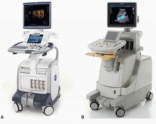 FIGURE 27.1. Examples of full-featured ultrasound systems. A, GE Logiq 9, photo courtesy of GE Healthcare. B, Philips iU22, photo courtesy of Philips Healthcare. |
Doppler Evaluation of Peripheral Arterial Pulses
A simple CW Doppler device can be used to assess peripheral arterial pulses during the physical examination (Fig. 27.3). As discussed in Chapter 7, CW Doppler signals do not allow for depth resolution and are not ideal for spectral waveform analysis; however, they can provide information on flow direction and produce signals that are suitable for audible interpretation. Experienced examiners can readily differentiate between the normal multiphasic peripheral arterial flow signal and the dampened, monophasic flow signal that is heard distal to a severe stenosis or occlusion. As described by Strandness and colleagues in 1967, a focal, high-pitched arterial signal represents the high-velocity jet within a stenosis.2 These qualitative findings can be used to establish the presence or absence of peripheral arterial disease and determine its approximate location and severity.
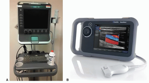 FIGURE 27.2. Examples of compact point of care ultrasound systems. A, SonoSite S-Series, photo courtesy of Amy Morris, MD. B, SonoSite NanoMaxx, photo courtesy of SonoSite, Inc. |
Ankle-Brachial Indices (without Doppler waveforms)
The measurement of systolic ankle pressures and calculation of the ankle-brachial indices is a simple quantitative method for assessing the status of the lower extremity arterial circulation (see Chapter 13). Although tibial artery flow waveforms are routinely recorded with the ankle pressures in a formal vascular laboratory examination, this step is not necessary at the point of care. Measurement of ankle and brachial systolic pressures and audible interpretation of the CW Doppler signals from the distal tibial arteries will provide a valuable physiologic assessment of the overall severity of lower extremity arterial occlusive disease. Figure 27.4 shows Dr. Strandness performing an ankle pressure measurement in the 1970s.
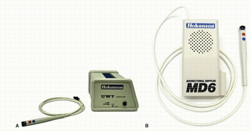 FIGURE 27.3. Continuous-wave (CW) Doppler instruments suitable for assessment of peripheral arterial pulses and measurement of ankle pressures. A, Portable Doppler. B, Pocket Doppler. Photos courtesy of D. E. Hokanson, Inc. |
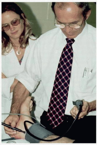 FIGURE 27.4. Dr. Strandness measuring an ankle pressure in the 1970s. |
COMPRESSION ULTRASOUND FOR DEEP VEIN THROMBOSIS
Screening for Proximal Lower Extremity Deep Vein Thrombosis
As discussed in Chapter 19, a complete lower extremity venous duplex examination performed in the vascular laboratory includes evaluation of multiple anatomic sites from the inferior vena cava to the tibial veins with B-mode imaging, pulsed Doppler spectral waveforms, and color Doppler. Although this comprehensive approach has become the standard test for diagnosis of acute deep vein thrombosis (DVT), a relatively quick 2-point compression ultrasound examination of the common femoral and popliteal veins has been advocated as a point of care screening test for acute proximal lower extremity DVT.9 In a randomized multicenter trial comparing whole-leg ultrasonography with the 2-point compression examination in 2465 outpatients, these two diagnostic strategies were found to be equivalent for the management of symptomatic patients with suspected DVT.10 The 2-point compression ultrasound examination has also been shown to compare favorably with CT venography for the diagnosis of femoropopliteal DVT with a sensitivity of 86% and specificity of 100%.11
The 2-point compression ultrasound examination relies completely on B-mode imaging; pulsed Doppler and color Doppler are not required. The common femoral and popliteal veins are imaged in a transverse view, and compressions are performed by downward pressure with the ultrasound probe, as illustrated in Chapter 19. After positioning the patient supine with the leg externally rotated, the common femoral vein is imaged from the inguinal ligament to the confluence of the deep femoral and femoral veins (Fig. 27.5). Next, the popliteal vein is imaged using a posterior approach from the adductor canal proximally to the confluence of the tibial veins distally (Fig. 27.6). The vein at both of these sites is compressed throughout its length in a transverse image. It is standard practice to point the indicator mark on the probe toward the right side of the patient for all transverse images, as shown in Figure 27.7, so the left side of the image will correspond to the patient’s right side—similar to the conventional orientation of a transverse image from a CT scan. With the posterior approach to the popliteal vein, the vein is typically closer to the transducer (skin line) than the artery (Fig. 27.8).
Stay updated, free articles. Join our Telegram channel

Full access? Get Clinical Tree


