Clinical classification
C0
No disease
C1
Telangiectasia
C2
Varicose veins
C3
Edema
C4a
Hemosiderin deposition, venous stasis dermatitis
C4b
Lipodermatosclerosis
C5
Healed venous ulcer
C6
Active/open venous ulcer
Etiologic classification
Ec
Congenital
Ep
Primary; idiopathic
Es
Secondary
Anatomic classification
As
Superficial
Ad
Deep
Ap
Perforator
Pathophysiologic classification
Pr
Reflux
Po
Obstruction
Pr,o
Reflux and obstruction in combination
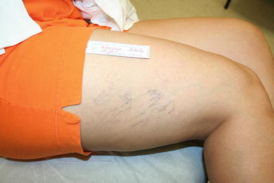
Fig. 12.1
Right leg showing telangiectasia, C1
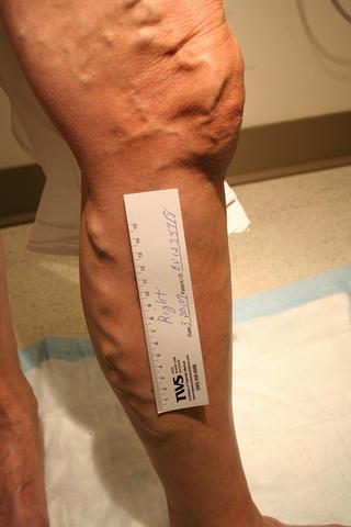
Fig. 12.2
Left leg varicose veins, C2
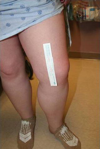
Fig. 12.3
Left leg edema, C3
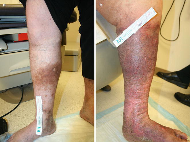
Fig. 12.4
(a) Right leg with hemosiderin deposits, C4a. (b) Left leg with lipodermatosclerosis, C4b
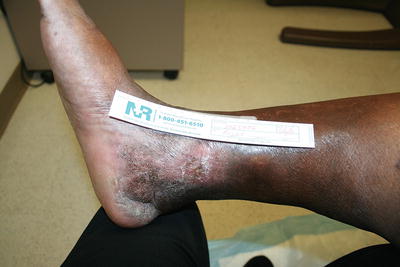
Fig. 12.5
Right leg with healed venous stasis ulcer, C5
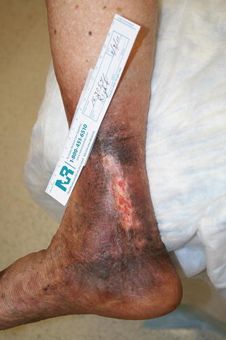
Fig. 12.6
Right leg with active venous stasis ulcer, C6
Table 12.2
Venous Clinical Severity Scoring (VCSS)

Diagnostic Evaluation: The Duplex Ultrasound Examination
Venous reflux refers to flow in the veins away from the heart and toward the periphery which is the opposite direction of the normal venous blood flow. This retrograde venous flow generates venous hypertension which can distend branch varicosities, trigger inflammation, and produce clinical symptoms such as leg fatigue, discomfort, and edema. Although this description oversimplifies the pathophysiology, there is little doubt that venous reflux plays an important role in varicose veins. All varicose vein treatment strategies attempt to eliminate or minimize the impact of venous reflux. The success of any therapy, therefore, requires a sensitive and practical test for detecting and characterizing venous reflux. Duplex ultrasonography meets these requirements and has become the gold standard imaging modality for evaluation of venous disease. It is a noninvasive test that can accurately assess all of the variables relevant to patients being evaluated for varicose veins including:
1.
Evidence of GSV and SSV reflux
2.
Severity and extent of superficial, perforator, and deep venous reflux
3.
Presence of variant tributaries of the superficial venous system
4.
Diameter of proximal, mid-, and distal GSV and SSV
5.
Patency of the deep venous system
Limitations of venous duplex ultrasound include its reliance on the sonographer’s technical skills and the inability to visualize veins in patients with extensive bandages, severe edema, and extremely obese body habitus.
Evaluating for reflux in the femoral veins and at the saphenofemoral junction should ideally be done with the patient standing for two reasons. First, standing dilates the lower extremity veins making them easier to identify and follow. Second, the upright position provokes reflux by increasing the hydrostatic pressure on the venous valves. Overall, the physiologic consequences of standing upright improve the sensitivity and specificity for detecting venous reflux. Conducting a duplex exam with the patient standing requires some safety precautions. A walker or fixed support bar allows patients to brace themselves in order to take weight off of the leg being examined. Keeping the knee in a slightly flexed position helps facilitate a complete exam. Before beginning, the patient should be instructed to communicate any feelings of dizziness that arise during the exam. In some patients, the overall atmosphere of the room coupled with the audible Doppler signals can elicit a fainting response. These symptoms tend to occur less frequently when the exam is performed silently. Patients who cannot tolerate standing during the exam can be evaluated on a stretcher placed in reverse Trendelenburg position.
The superficial axial veins, that is, the GSV and SSV, should be mapped and interrogated with sequential compression throughout their course. The GSV resides in a fascial sheath with sonographically distinct anterior and posterior borders. When compressed the normal GSV appears as a winking eye within the sheath [5]. Incomplete compression indicates acute thrombus or chronic phlebitis if the image is associated with thickened walls or intraluminal septae. Vein segments visualized outside of the saphenous sheath are accessory vein branches, not the axial vein, and anomalous venous anatomy or branching should be noted. The SSV appears in a triangular sheath created by the two heads of the gastrocnemius and the surrounding fascia.
Reflux testing employs provocative maneuvers designed to elicit retrograde venous flow. The Valsalva maneuver generates retrograde venous flow in the proximal GSV. With the transducer positioned to visualize the saphenofemoral junction, the patient takes a deep breath and bears down with a closed epiglottis. Color flow imaging demonstrates reversed flow in the proximal GSV. The exact duration of retrograde flow can then be determined by reviewing the spectral analysis of the venous waveform recorded during the Valsalva maneuver. Venous reflux is defined as prolonged retrograde flow lasting longer than 0.5 s (500 ms). The Valsalva test for reflux can only be used to evaluate the saphenofemoral junction since a competent valve will interrupt the transmission of pressure distally [6].
Evaluating for reflux in the more distal veins of the lower extremity employs a series of compression and release maneuvers. After distal compression and rapid release, venous flow reverses until contact with a competent venous valve. In perforator veins, a cutoff time of 350 ms is used to define venous reflux, while 1,000 ms is used for the femoral and popliteal veins (Table 12.3). To be effective, the compression and release of the thigh and calf should be done quickly and sharply. Automated pressure cuffs can substitute for manual compression and offer a more standardized, albeit more cumbersome, exam [6].
Table 12.3
Criteria for reflux
Vein | Criteria |
|---|---|
Femoral, popliteal tibial | Greater than 1,000 ms |
Great and small saphenous (GSV, SSV) | Greater than 500 ms |
Perforating veins | Greater than 350 ms (diameter greater than 3.5 mm) |
In addition to evaluating for reflux, duplex examination also provides essential information regarding superficial venous anatomy, patency, and diameter. Duplicated veins and accessory tributaries occur relatively frequently in the lower extremity. More than 18 % of patients have duplicated saphenous systems, and other veins may be hypoplastic or aplastic. The diagnostic duplex study should actively search for and characterize duplicated and accessory veins. In some cases, reflux may occur only in an accessory venous tributary, while the GSV remains uninvolved. Knowledge of these anatomic variants allows practitioners to direct venous interventions at only those segments which demonstrate venous reflux [7].
The duplex ultrasound exam should also assess the patency of the superficial and deep veins because the presence of venous thrombosis or obstruction can limit interventional treatment options. Complete venous wall apposition during compression confirms patency. A dilated vein that fails to compress indicates acute thrombosis, while thickened vein walls and altered inhomogeneous color flow suggest chronic thrombosis or partial recanalization. Acute or chronic obstruction of the GSV or SSV may prevent advancement of the venous stripper or ablation catheter precluding surgical vein stripping or endovenous ablation, respectively. Likewise, obstruction of the deep venous system represents a relative contraindication to interventions on the superficial venous system. In these cases, the superficial axial veins should be preserved because they may be acting as important venous collaterals providing venous outflow for the lower extremity.
The diameter of the GSV helps determine the presence of venous reflux. Diameters larger than 7.3 mm at the saphenofemoral junction, 6 mm in the midthigh, and 4 mm in the midcalf are predictive of an incompetent GSV. In contrast, diameters smaller than 5.5 mm, 3 mm, and 2 mm at the same respective levels are associated with GSV competence. Vein diameter and distance from the skin also play a role in planning for endovenous ablation procedures. These variables help determine what energy level will ablate the vein without risking thermal injury to the skin. Ultrasound diameter measurements of the saphenous and accessory veins should be conducted with the patient supine since ablation procedures are performed in that position. The GSV 3–4 cm below the knee deserves particular attention because it is the most common entry point for percutaneous access and it must be of adequate caliber to accept the therapeutic catheters.
Noninterventional Management
The initial management of varicose veins usually involves noninterventional therapy consisting of compression stockings, lower extremity elevation, exercise, and anti-inflammatory medication as needed. Compression stockings exert graduated pressure on the lower extremity which keeps varicose veins from becoming distended during prolonged standing, walking, and sitting with the legs in a dependent position. This extrinsic compression may provide symptomatic relief by minimizing the inflammatory response triggered by varicose vein engorgement and lower extremity edema. Compression stockings come in a variety of lengths (knee high, thigh high, pantyhose) and various pressure ranges: 8–15 mmHg, 15–20 mmHg, 20–30 mmHg, 30–40 mmHg, and greater than 40 mmHg. An adequate trial of noninterventional therapy requires use of compression stockings with a minimum pressure of 20–30 mmHg.
Although compression stockings effectively control the symptoms of varicose veins, they cannot work if they are not worn regularly. Patients should be instructed to put the stockings on as soon as they get out of bed in the morning when leg edema and varicose vein distention are least prominent. They should wear the stockings all day while performing their activities of daily living and remove them in the evening prior to bed. Some patients lack the physical strength or manual dexterity to pull on their stockings. Advanced age, severe musculoskeletal disease, and high body mass index can render stocking use nearly impossible. In addition, patients who have concomitant arterial occlusive disease (ABI less than 0.5) should not wear compression stockings to avoid compromising the tenuous arterial perfusion. Physicians can improve patient compliance by emphasizing the advantages of compression stockings including their noninvasive nature, excellent safety profile, and ability to improve symptoms with use. Below-knee stockings are the easiest to wear and should be prescribed regularly unless patients have varicosities around the popliteal fossa. Patients must be measured by a certified stocking fitter who can act as a useful resource to help patients understand proper stocking application. Compliance generally improves with stocking education.
Lower extremity elevation allows passive emptying of the axial and tributary veins and provides relief of symptoms associated with ambulatory venous hypertension. Patients are instructed to keep their “toes above the nose” for 15 min twice a day. Despite being brief in duration, these intervals of lower extremity venous decompression can alleviate some of the symptoms associated with varicose veins.
Exercise plays an essential role in noninterventional management of varicose veins and superficial venous insufficiency. The specific type of exercise is not important as long as it involves activation of the gastrocnemius and soleus muscles. Calf muscle contraction pumps venous blood from the GSV and SSV of the superficial system into the femoral and popliteal veins of the deep system. The lower extremity venous pump also augments venous blood flow from the distal leg into the proximal thigh and ultimately increases venous outflow into the iliac veins and inferior vena cava. Overall, exercise decreases lower extremity venous pressure [8]. Although not required, some patients report that wearing compression stockings during exercise provides additional symptomatic relief.
Medical therapy for venous disease primarily involves anti-inflammatory medications to relieve symptoms associated with varicose veins and superficial phlebitis. Outside of the United States, venoactive agents are used more regularly to treat symptomatic venous insufficiency. Studies involving horse chestnut extract, gamma-benzopyrenes, flavinoids, and pine bark extract have reported a decrease in leg edema. Although these small studies had promising results, a recent Cochrane review concluded that there was insufficient evidence to support widespread use of venoactive agents. In patients with advanced chronic venous insufficiency, flavinoids (MPFF) or pentoxifylline may accelerate ulcer healing, and the Society of Vascular Surgery/American Venous Forum (SVS/AVF) Guidelines Committee recommends using one of these agents as adjuvant therapy to compression (Grade 2B) [9].
Treatment
Indications for Intervention
The initial management of symptomatic varicose veins consists of a trial of conservative measures including compression stockings, anti-inflammatory medication, and leg elevation. After 3 months, patients should be reevaluated to determine the success of noninterventional therapy. If the patient’s symptoms persist despite adequate compliance, interventional treatment options should be considered. Patients who present with complications related to varicose veins such as superficial thrombophlebitis or bleeding can often forego a trial of conservative treatment and be considered for immediate intervention.
All interventions for varicose veins share the same treatment objective: to remove branch varicosities and eliminate superficial venous reflux. Various forms of phlebectomy and sclerotherapy physically remove or close off branch varicosities, thereby abolishing the source of symptoms and the most obvious physical manifestation of the disease. Procedures directed at superficial venous reflux focus on the unseen source of venous hypertension and varicose veins. Surgical stripping and percutaneous ablation of the GSV are techniques designed to reduce venous hypertension by eliminating superficial venous reflux. In addition to providing symptomatic relief, successful vein stripping or ablation procedures also decrease the rate of recurrent varicose veins.
Surgical Vein Stripping
Surgical techniques for eliminating superficial venous reflux started to develop over 100 years ago. Keller introduced saphenous vein invagination and stripping, while Mayo pioneered the use of an external stripper to remove the saphenous vein. Babcock described stripping the saphenous vein intraluminally from the ankle to groin. High ligation of the GSV briefly gained popularity as a method for treating venous reflux without removing the GSV. Unfortunately, ligation usually fails to eliminate venous reflux, and it has been largely abandoned as an isolated treatment for superficial venous incompetence. Modern surgical treatment for superficial venous reflux involves high ligation and stripping of the GSV from the knee to the groin. This method of GSV removal minimizes the risk of nerve injury compared to stripping that extends to the ankle.
High ligation and vein stripping usually requires general or spinal anesthesia. A transverse or oblique groin incision is made just medial to the femoral artery pulse and inferior to the inguinal crease. Sharp dissection allows identification of the proximal GSV and other venous tributaries which can be ligated and divided. A brief exploration to identify the presence of a duplicate saphenous system should be performed. The GSV can then be brought up into the surgical field with gentle traction on the saphenofemoral junction. This maneuver affords further visualization of any missed tributaries that require ligation. The GSV should be ligated with a nonabsorbable suture and transected near its confluence with the femoral vein.
Stay updated, free articles. Join our Telegram channel

Full access? Get Clinical Tree


