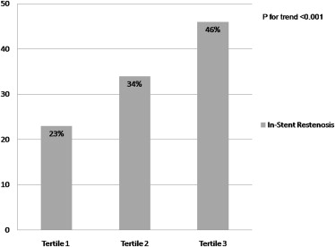
Serum uric acid (SUA) level is known as a significant predictor for cardiovascular diseases, partly through increased inflammatory response and smooth muscle cell proliferation. Inflammation and smooth muscle cells play a crucial role in the pathogenesis of in-stent restenosis (ISR). However, the relation between SUA and ISR has not been studied. The aim of the present study was to investigate the predictive value of preprocedural SUA on the development of ISR in patients who undergo coronary bare-metal stent implantation. Clinical, biochemical, and angiographic data from 708 consecutive patients (mean age 60.3 ± 9.3 years, 71% men) who had undergone bare-metal stent implantation and additional control coronary angiography for stable or unstable angina pectoris were analyzed. Patients were divided into tertiles on the basis of preprocedural SUA levels. Stent restenosis was observed in 54 patients (23%) in the lowest tertile, in 79 (34%) in the middle tertile, and in 109 (46%) in the highest tertile (p <0.001). Using multiple logistic regression analysis, diabetes mellitus, smoking, high-density lipoprotein cholesterol, stent length, C-reactive protein level, and preprocedural SUA level emerged as independent predictors of ISR. On receiver-operating characteristics curve analysis, SUA level >5.5 mg/dl had 75% sensitivity and 71% specificity (area under the curve 0.784, p <0.001) in predicting ISR. In conclusion, higher preprocedural SUA is a powerful and independent predictor of bare-metal stent restenosis in patients with stable and unstable angina pectoris.
Despite considerable technological evolution in percutaneous coronary intervention (PCI), in-stent restenosis (ISR) has continued to be a major drawback, and great efforts have been made to resolve this vexing problem. Biologic responses related to PCI-induced vascular injury are characterized by the sequence of inflammation, granulation, extracellular matrix remodeling, and smooth muscle cell (SMC) proliferation and migration, which leads to neointimal hyperplasia and restenosis. Besides technical and mechanical conditions associated with PCI, inflammatory status before and after stent implantation is a significant risk factor for ISR. It has been shown that serum uric acid (SUA) is linked to the presence, severity, and progression of atherosclerosis through proinflammatory properties and stimulation of vascular SMC proliferation. Although various proinflammatory biomarkers have been used in clinical practice to evaluate the association of inflammatory status with ISR, to our knowledge, no study has investigated any possible association between preprocedural SUA level and ISR. The aim of the present study was to evaluate the association of SUA level before successful bare-metal stent (BMS) implantation in predicting ISR in patients with stable and unstable angina pectoris.
Methods
We analyzed clinical, laboratory, and angiographic data from consecutive patients who underwent successful BMS implantation from January 2008 to December 2010 at Türkiye Yüksek Ihtisas Educational and Research Hospital (Ankara, Turkey). The inclusion criteria were as follows: (1) stable or unstable angina, (2) coronary angiography showing de novo lesions without a history of PCI, and (3) coronary stent implantation. For patients’ data, we gained access retrospectively to the data at the time of interest, when the patients underwent BMS implantation after control coronary angiography performed because of clinical indications, including symptoms of angina and abnormal noninvasive test results (either treadmill exercise tests or myocardial perfusion scintigraphy), thus recalling clinical, angiographic, and laboratory characteristics at that time. As such, we were able to collect data from 770 patients. Patients with unstable angina pectoris were identified according to the definition of Braunwald. As part of our preprocedural protocol, SUA level was available before coronary angiography in all patients. Patients were excluded from the analysis if they had clinical evidence of neoplastic diseases (n = 3), renal dysfunction (glomerular filtration rate <90 ml/min/1.73 m 2 ; n = 35), hepatic and hemolytic disorders (n = 2), chronic inflammatory disease (n = 5), any active infectious disease (n = 3), alcohol consumption (n = 6), vitamin use (including vitamin C, niacin, and folate; n = 3), major adverse events during follow-up (n = 3), and SUA-lowering medications such as allopurinol (n = 2), leaving 708 patients to be included in the study for analysis.
Patients’ clinical and demographic characteristics, encompassing age, gender, history of arterial hypertension, diabetes mellitus, tobacco use, family history of coronary artery disease, the left ventricular ejection fraction, and medications used, were noted. In addition, serum levels of fasting blood glucose, creatinine, C-reactive protein (CRP), and lipid panel, including total cholesterol, low-density lipoprotein cholesterol, high-density lipoprotein cholesterol, and triglyceride levels, were also recorded. The local ethics committee approved the study protocol.
All laboratory data were obtained from venous blood samples up to 6 hours before stent implantation. Lipid profile, glucose, creatinine, and CRP levels were measured according to standard methods. SUA levels were measured using an enzymatic colorimetric method (Cobas Integra Uric Acid Cassette; Roche Diagnostics, Indianapolis, Indiana) on an autoanalyzer (Cobas Integra 400; Roche Diagnostics).
Coronary interventions were performed according to current practice guidelines and recorded in digital storage for further analysis. The degree of coronary stenosis was visually estimated by experienced interventional cardiologists. Luminal narrowing >50% in a major subepicardial vessel (left anterior descending, left circumflex, or right coronary artery) was defined as significant stenosis. Each patient received aspirin plus clopidogrel (loading dose 300 or 600 mg) before or during coronary intervention. Unfractionated heparin 100 U/kg was administered at the beginning of the procedure to keep the activated clotting time >200 seconds. The access site for PCI was at the physician’s preference (femoral or radial). Use of glycoprotein IIb/IIIa inhibitors and predilatation or postdilatation after stent implantation of the lesion was at the operator’s discretion. Successful PCI was defined as a <20% decrease in diameter stenosis and residual stenosis <5% in diameter, with final Thrombolysis In Myocardial Infarction (TIMI) grade 3 flow without any major complications. After stent placement, only 1 month of clopidogrel was used, and aspirin was used indefinitely. During routine clinical follow-up, coronary angiography was performed for clinical indications secondarily in patients with stable or unstable angina pectoris. Control coronary angiograms were recorded using the Judkins technique and interpreted by 2 independent cardiologists who were blinded to patients’ data. The evaluation of stenosis was carried out using the conventional visual assessment technique. Stent restenosis was accepted as narrowing >50% in a vessel of otherwise normal diameter, including 5 mm proximal and distal to the stent edge, according to results of control coronary angiography. Intra- and interobserver variability of stent restenosis analysis was minimal in a representative subset of 80 patients. The interpretations of the 2 investigators on the presence or absence of ISR agreed in 96% (77 of 80) and 97.5% (78 of 80), respectively. Intraobserver variability was assessed by 1 investigator. The 2 readings were concordant for the presence or absence ISR in 97.5% (78 of 80) and 97.5% (78 of 80), respectively.
Analyses were performed using SPSS version 13.0 (SPSS, Inc., Chicago, Illinois). Continuous data are presented as medians and interquartile ranges or as mean ± SD. To test the distribution pattern, the Kolmogorov-Smirnov test was used. The study population was assigned into tertiles on the basis of preprocedural SUA level. Comparisons of multiple mean values were carried out using Kruskal-Wallis tests or analysis of variance as appropriate. Categorical variables were summarized as percentages and compared using chi-square tests. Spearman’s correlation coefficient was computed to examine the association between 2 continuous variables. Effects of different variables on ISR were calculated in univariate analysis for each. Variables for which the unadjusted p values were <0.10 in logistic regression analysis were identified as potential risk markers and included in the full model. We reduced the model using stepwise multivariate logistic regression analyses and eliminated potential risk markers using likelihood ratio tests. A p value <0.05 was considered statistically significant, and the confidence interval was 95%. Analysis was performed in 2 models. Preprocedural SUA level was assumed to be a continuous variable in the first model and a categorical variable in the second model. An exploratory evaluation of additional cut points was performed using receiver-operating characteristic curve analysis. A p value <0.05 was considered statistically significant.
Results
The baseline clinical and procedural characteristics of the study population are summarized in Table 1 . A total of 708 patients (mean age 60.3 ± 9.3 years, 71% men) were grouped into tertiles according to preprocedural SUA level. The mean period between the 2 coronary angiographic studies for all study population was 20.4 ± 4.5 months.
| Variable | SUA Level (mg/dl) | p Value | ||
|---|---|---|---|---|
| 3.9–4.9 (n = 236) | 5.2–5.9 (n = 236) | 6.3–7.4 (n = 236) | ||
| Age (yrs) | 60.1 ± 9.3 | 59.5 ± 9.2 | 61.2 ± 10.1 | 0.49 |
| Men | 174 (74%) | 164 (69%) | 165 (70%) | 0.36 |
| Body mass index (kg/m 2 ) | 27.5 ± 4.0 | 28.2 ± 5.3 | 28.2 ± 4.4 | 0.44 |
| Diabetes mellitus | 54 (23%) | 71 (30%) | 86 (36%) | 0.001 |
| Current smokers | 81 (34%) | 93 (39%) | 114 (48%) | 0.002 |
| Hypertension | 132 (56%) | 125 (53%) | 134 (57%) | 0.85 |
| Reason for stent implantation | ||||
| Stable angina pectoris | 165 (71%) | 155 (66%) | 147 (61%) | 0.08 |
| Unstable angina pectoris | 71 (30%) | 81 (34%) | 89 (38%) | 0.08 |
| Number of coronary arteries narrowed | ||||
| 1 | 47 (20%) | 47 (20%) | 57 (24%) | 0.26 |
| 2 | 189 (80%) | 189 (80%) | 179 (76%) | 0.26 |
| Target coronary artery | ||||
| Left anterior descending | 124 (53%) | 100 (42%) | 104 (44%) | 0.06 |
| Right | 51 (22%) | 83 (35%) | 76 (32%) | 0.012 |
| Left circumflex | 61 (26%) | 53 (22%) | 56 (24%) | 0.59 |
| Stent diameter (mm) | 3 (2.75–3) | 3 (2.5–3) | 3.25 (2.75–3) | 0.44 |
| Stent length (mm) | 15 (12.5–18) | 15.5 (13–17.5) | 16.5 (13.5–18) | 0.33 |
| In-hospital medical treatment | ||||
| β blockers | 82% | 82% | 79% | 0.83 |
| Angiotensin-converting enzyme inhibitors | 75% | 78% | 77% | 0.82 |
| Calcium channel blockers | 4% | 4% | 3% | 0.84 |
| Angiotensin receptor blockers | 5% | 6% | 5% | 0.81 |
| Statins | 88% | 89% | 88% | 0.88 |
| Ejection fraction (%) | 62.1 ± 3.7 | 61.4 ± 4.0 | 60.4 ± 3.7 | 0.47 |
| Fasting glucose (mg/dl) | 103 (90–129) | 107 (93–139) | 114 (94–143) | 0.04 |
| High-density lipoprotein-cholesterol (mg/dl) | 42 (34–46) | 39 (33–46) | 34 (30–42) | 0.01 |
| Low-density lipoprotein-cholesterol (mg/dl) | 108 (82–135) | 107 (84–139) | 110 (85–1,339) | 0.75 |
| Triglycerides (mg/dl) | 133 (99–201) | 137 (102–193) | 144 (102–204) | 0.19 |
| Serum creatinine (mg/dl) | 0.8 (0.7–0.9) | 0.9 (0.8–1.0) | 1.0 (0.9–1.1) | 0.001 |
| CRP (mg/dl) | 1.7 (0.85–3.25) | 3.05 (1.55–6.55) | 5.95 (3.85–15.5) | <0.001 |
| Hemoglobin (g/L) | 14.4 ± 2.1 | 14.0 ± 1.8 | 13.7 ± 1.7 | 0.02 |
| White blood cell count (×10 9 /L) | 7.8 (6.9–9.3) | 8.2 (7.3–9.7) | 8.4 (7.0–9.9) | 0.27 |
| Platelet count (×10 9 /L) | 255.8 ± 30.2 | 249.6 ± 35.7 | 256.8 ± 34.2 | 0.53 |
| Time between the 2 coronary angiographic studies (mo) | 21.2 ± 4.3 | 20.5 ± 5.4 | 19.4 ± 4.1 | 0.21 |
| ISR | 54 (23%) | 79 (34%) | 109 (46%) | <0.001 |
Patients in tertile 3 had significantly higher rates of ISR compared with those in tertiles 1 and 2 (p <0.001; Figure 1 ). Likewise, higher SUA levels before stent implantation were found to be associated with increased ISR by logistic regression analysis. A positive correlation was found between SUA and CRP level (r = 0.412, p <0.001). Assuming SUA level as a continuous (model 1) or categorical (model 2) variable in multiple logistic regression analysis, diabetes mellitus, smoking, high-density lipoprotein cholesterol, stent length, CRP, and SUA level emerged as independent predictors of ISR ( Table 2 ). Receiver-operating characteristic curve analysis was used to explore the relation between preprocedural SUA and ISR. The area under the curve was 0.784 (95% confidence interval 0.75 to 0.91, p <0.001). Preprocedural SUA with a cut-off level of >5.5 mg/dl predicted ISR with sensitivity of 75% and specificity of 71% ( Figure 2 ). Also, mean SUA level was found to be significantly increased in the ISR group in patients with stable angina pectoris (5.7 ± 1.1 vs 4.8 ± 1.1 mg/dl, p <0.001) and unstable angina pectoris (5.9 ± 1.3 vs 4.9 ± 1.2 mg/dl, p <0.001).

| Variable | Univariate | Multivariate | ||
|---|---|---|---|---|
| OR (95% CI) | p Value | OR (95% CI) | p Value | |
| Model 1: SUA level as a continuous variable | ||||
| Age | 1.05 (0.94–1.16) | 0.42 | — | — |
| Diabetes mellitus | 1.31 (1.13–1.50) | <0.001 | 1.20 (1.08–1.31) | <0.001 |
| Smoker | 1.53 (1.09–1.97) | <0.001 | 1.62 (1.33–1.92) | <0.001 |
| High-density lipoprotein | 0.90 (0.84–0.95) | 0.001 | 0.92 (0.86–0.98) | 0.02 |
| Low-density lipoprotein | 1.07 (0.95–1.19) | 0.12 | — | — |
| SUA | 1.22 (1.10–1.35) | 0.001 | 1.07 (1.03–1.12) | 0.003 |
| Triglycerides | 1.03 (0.84–1.20) | 0.48 | — | — |
| Serum creatinine | 1.09 (0.94–1.23) | 0.14 | — | — |
| CRP | 1.30 (1.24–1.37) | <0.001 | 1.09 (1.06–1.12) | 0.001 |
| Hemoglobin | 0.97 (0.92–1.03) | 0.25 | — | — |
| Stent length | 1.23 (1.11–1.36) | 0.001 | 1.31 (1.15–1.47) | 0.002 |
| Stent diameter | 1.03 (0.95–1.11) | 0.49 | — | — |
| Left ventricular ejection fraction | 1.01 (0.97–1.05) | 0.36 | — | — |
| Time between the 2 coronary angiographic studies | 0.92 (0.88–0.96) | 0.01 | 0.97 (0.92–1.03) | 0.10 |
| Model 2: SUA level as a categorical variable | ||||
| Age | 1.05 (0.94–1.16) | 0.42 | — | — |
| Diabetes mellitus | 1.31 (1.13–1.50) | <0.001 | 1.29 (1.11–1.46) | <0.001 |
| Smoker | 1.53 (1.09–1.97) | <0.001 | 1.60 (1.24–1.96) | <0.001 |
| High-density lipoprotein | 0.90 (0.84–0.95) | 0.001 | 0.93 (0.88–0.98) | 0.03 |
| Low-density lipoprotein | 1.07 (0.95–1.19) | 0.12 | — | — |
| SUA (>5.5 mg/dl) | 1.45 (1.21–1.69) | <0.001 | 1.24 (1.13–1.35) | 0.001 |
| Triglycerides | 1.03 (0.84–1.20) | 0.48 | — | — |
| Serum creatinine | 1.09 (0.94–1.23) | 0.14 | — | — |
| CRP | 1.30 (1.24–1.37) | <0.001 | 1.11 (1.04–1.18) | <0.001 |
| Hemoglobin | 0.97 (0.92–1.03) | 0.25 | — | — |
| Stent length | 1.23 (1.11–1.36) | 0.001 | 1.28 (1.12–1.45) | 0.01 |
| Stent diameter | 1.03 (0.95–1.11) | 0.49 | — | — |
| Left ventricular ejection fraction | 1.01 (0.97–1.05) | 0.36 | — | — |
| Time between the 2 coronary angiographic studies | 0.92 (0.88–0.96) | 0.01 | 0.98 (0.94–1.02) | 0.15 |
Stay updated, free articles. Join our Telegram channel

Full access? Get Clinical Tree


