Use of Diagnostic Testing

3 Use of Diagnostic Testing


The physician confronted with a patient with suspected cardiovascular disease has a multitude of tests available to provide diagnostic and prognostic information. Chapters 4 through 10 describe the various modalities for diagnosing cardiovascular diseases. This chapter focuses on the selection of the most appropriate tests for individual patients.


Generally, the available cardiovascular diagnostic tests can be divided into two categories: Tests that assess anatomy and tests that assess function. These categories are merging, as tests once used solely for anatomic purposes are modified to also assess function. The choice of test depends not only on the question being asked but also on the cost-effectiveness and predictive value of the test and the relative value of anatomic versus functional information. An anatomic assessment (using a test validated by comparison with coronary angiography) may be useful in some settings, but it does not eliminate the need for a functional assessment, which may be even more predictive of a patient’s prognosis and need for further intervention.


New imaging techniques must be carefully evaluated for accuracy, ability to provide the needed information, and cost-effectiveness compared with existing methods of obtaining similar information. It should be noted that the initial description of the sensitivity and specificity of a diagnostic test may overestimate what can be achieved in practice. Initial publications usually describe the assessment of a diagnostic test under rigorous conditions by experienced operators in a highly selected population. The true measure of a test is its ability to produce reliable information in a typical clinical environment.


This chapter reviews the available tests that most frequently provide diagnostic and prognostic information in the evaluation of patients with suspected cardiovascular disease. As with all diagnostic tests, the pre-test probability of disease must be considered carefully, both in choosing the most appropriate test and in its interpretation.


Diagnostic Tests


Electrocardiography


The resting ECG is the most frequently performed investigation in evaluating patients with cardiovascular disease (see Chapter 4). Electrocardiography is a highly versatile diagnostic test, providing information on a broad spectrum of clinical conditions, ranging from metabolic disturbances (e.g., hypo- and hyperkalemia) and pharmacologic toxicity to ischemic heart disease (e.g., acute myocardial infarction [MI], unstable angina), arrhythmia, and pericardial disease (see Chapter 4). With such versatility, this simple-to-perform test is cost effective.


In the investigation of suspected or known arrhythmias, Holter electrocardiographic monitoring augments the resting ECG by allowing correlation of patient symptoms to the rhythm disturbance and the subsequent monitoring of the patient’s response to treatment. This can be in the form of a continuous 24- to 72-hour monitor, a patient-activated event monitor worn for 1 to 4 weeks, or a subcutaneous Reveal device (up to 2 years). Continuous ST-segment monitoring also collects prognostic data on patients who have had a coronary event.


Exercise ECG is a relatively inexpensive investigation used in the diagnosis and management of coronary artery disease (CAD). However, with a sensitivity of approximately 67% and a specificity of 84% for the detection of significant CAD in an optimal setting (and much lower accuracy reported in other settings), the main value of exercise ECG lies in excluding CAD in patients who have a moderate or low pre-test probability of significant coronary stenoses. The risk of false-negative results in patients with a high pre-test probability of CAD is relatively high; these patients should be referred for a more sensitive test such as coronary angiography.


Biochemical Markers


Serum troponin T and I are highly sensitive and specific markers for myocardial injury that are widely accepted as the standard biomarkers for the diagnosis of MI. Elevated troponin levels predict mortality in acute coronary syndromes as well as other diseases, including heart failure, renal failure, and sepsis. In acute MI, serum troponin levels rise after 2 to 3 hours, become detectable in the bloodstream at 6 to 12 hours, and remain elevated for up to 14 days. Caution interpreting positive troponin results should be used, however, because of the wide range of nonischemic cardiac and noncardiac conditions that can cause elevated serum concentrations. These conditions are numerous and include tachyarrythmia, myocarditis, direct current cardioversion, renal failure, sepsis, pulmonary embolism, and stroke. The other main caution in interpretation is an understanding of the details of the test used locally; the many commercially available assays have different upper limits of normal.


Brain natriuretic peptide and its co-secreted N-terminal fragment are useful in the diagnosis of acute heart failure in an emergency department and management of chronic heart failure in a primary care setting. They may be useful to establish prognosis in heart failure, in that both markers are typically higher in patients with worse outcomes. Although they are highly sensitive and therefore have very few false-negative results, they unfortunately lack the specificity needed to exclude false-positive results and are often therefore used as a “rule out” test for heart failure. Serum levels are elevated in patients with renal failure, because both peptides are renally excreted. Results should be interpreted in the clinical context, and positive results should invariably be followed by functional imaging such as an echocardiogram to formally assess cardiac function.


Echocardiography


Echocardiography provides a versatile and cost-effective method for assessing cardiac anatomy and function (see Chapter 6). The greatest values of echocardiography are the capacity for simultaneous assessment of valvular, pericardial, myocardial, and extracardiac abnormalities. Because complex image processing is not needed, the results of the study are immediately available to the experienced echocardiographer. In addition, it is possible to perform echocardiography on critically ill patients who cannot be moved, or in other circumstances when a portable test is preferable. For these reasons, echocardiography is the preferred screening imaging test for further assessing suspected myocardial dysfunction. Moreover, the use of Doppler echocardiography (Doppler) to measure flow allows the measurement of peak velocity across valves, the mapping of regurgitant jets, the estimation of pulmonary artery pressures, and the detection of shunts (e.g., ventricular and atrial septal defects). The severity of valvular heart disease and its contribution to the clinical presentation can be determined immediately. For patients with chest pain, congestive heart failure, or arrhythmias, echocardiography provides a rapid means of determining underlying cardiovascular function.


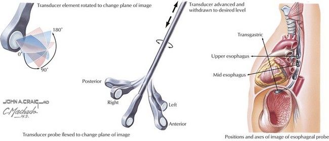
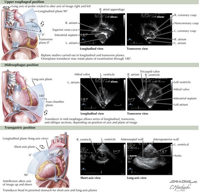
Transesophageal echocardiography adds to the sensitivity of transthoracic echocardiography, because views of the heart are not impeded by artifact related to the lungs or chest wall (Figs. 3-1 and 3-2). In addition, transesophageal echocardiography allows visualization of structures that are usually not well seen by transthoracic echocardiography (e.g., the left atrial appendage). The development of transesophageal echocardiography has also been an important advance in the management of patients who are undergoing cardiothoracic surgery, providing information on left ventricular (LV) function and the success of valvular repair. In addition, transesophageal echocardiography may allow a more accurate determination of valvular dysfunction and assessment for bacterial endocarditis, intracardiac thromboses, or both.


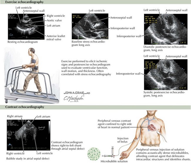
In addition to its usefulness in assessing valvular heart disease, echocardiography provides information on regional wall motion abnormalities suggestive of myocardial ischemia or necrosis in patients with CAD. The addition of pharmacologic or exercise-induced stress to detect inducible ischemia provides increased sensitivity and specificity compared with ECG exercise testing (Fig. 3-3, upper panel). In 21 studies, the sensitivity of exercise stress echocardiography averaged 84% (range 71% to 97%) and the specificity averaged 86% (range 64% to 100%). The use of echocardiography can be limited by technical considerations, including an inability to obtain diagnostic images in some patients (an estimated 15%). Stress echocardiography is indicated for individuals who have an intermediate prior probability of CAD and for individuals with abnormal ECGs or who are prescribed medications that can cause ECG abnormalities with stress (such as digoxin). In either of these cases the predictive value of exercise ECG is substantially reduced, justifying the use of an imaging technique during stress.


Stay updated, free articles. Join our Telegram channel

Jun 12, 2016 | Posted by in CARDIOLOGY | Comments Off on Use of Diagnostic Testing

Full access? Get Clinical Tree

Get Clinical Tree app for offline access