Upper Respiratory Complaints
A 65-year-old woman presents to her primary care physician with upper respiratory complaints. She later develops progressively worsening dyspnea on exertion and cough.
She is treated with antibiotics with limited resolution of her symptoms.
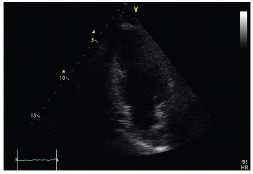
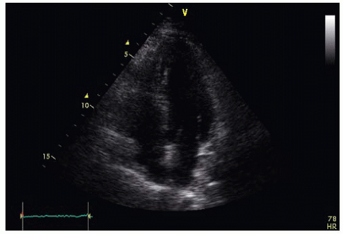
Transthoracic echocardiography (TTE) is performed. Figures 52-1 and 52-2 and Videos 52-1 and 52-2 show the parasternal long axis and apical four-chamber views, and no abnormalities were seen.
Two months later, the patient is hospitalized after presenting to her ophthalmologist, complaining of loss of vision in her right eye. Retinal examination reveals evidence of an embolus to the right eye.
Brain magnetic resonance imaging reveals evidence of cerebral infarction localized to the caudate nucleus and both cerebellar hemispheres.
TTE is repeated; Figure 52-3 and Video 52-3 show apical four-chamber view.
 Figure 52-2.
Stay updated, free articles. Join our Telegram channel
Full access? Get Clinical Tree
 Get Clinical Tree app for offline access

|
