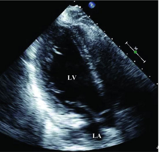
Figure 81.2 Transthoracic echocardiography: apical long-axis view shows the ventricular anterior septal and posterior wall are normal in thickness. LA, left atrium; LV, left ventricle.
The left atrium and right atrium are normal in size and structure. Mild tricuspid regurgitation is detected. The tricuspid regurgitant velocity is 2.93 m/sec, and the estimated pulmonary artery systolic pressure is moderately elevated (44.3 mmHg).
Pulse Doppler from mitral valve, pulse tissue Doppler from mitral annulus septal corner and color M-mode recordings show E/E′ = 15.4 and the slope of color M-mode = 44.5 cm/sec (Figure 81.3). These results indicate a 25-year-old young man with diastolic dysfunction.


