, Marilyn J. Siegel2, Tomasz Miszalski-Jamka3, 4 and Robert Pelberg1
(1)
The Christ Hospital Heart and Vascular Center of Greater Cincinnati, The Lindner Center for Research and Education, Cincinnati, OH, USA
(2)
Mallinckrodt Institute of Radiology, Washington University School of Medicine, St. Louis, Missouri, USA
(3)
Department of Clinical Radiology and Imaging Diagnostics, 4th Military Hospital, Wrocław, Poland
(4)
Center for Diagnosis Prevention and Telemedicine, John Paul II Hospital, Kraków, Poland
Abstract
Uhl anomaly is a very rare condition characterized by complete or partial absence of the right ventricular myocardium, which is replaced by fibroelastic tissue [1].
Uhl anomaly is a very rare condition characterized by complete or partial absence of the right ventricular myocardium, which is replaced by fibroelastic tissue [1].
The true incidence of Uhl anomaly is unknown, but less than 100 confirmed cases have been described in the literature [2]. The cause is thought to be a high apoptotic activity, which begins during the perinatal period or early in infancy, leading to destruction of the right ventricular wall [2, 3]. Histologic examination reveals partial or total absence of the myocardium of the parietal wall of the right ventricle and direct apposition of the opposing endocardial and epicardial surfaces [4, 5]. This leads to thinning of the right ventricular free wall. Uhl anomaly usually presents in neonates or infants as right-sided heart failure. Patients rarely survive to adulthood. No effective treatment other than heart transplant has been shown to improve survival.
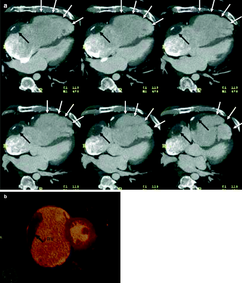
Computed tomography (CT) findings include an extremely thin-walled right ventricle with complete or partial absence of the right ventricular free-wall myocardium and a paucity of trabeculations (Figs. 8.1 and 8.2) [6, 7]. The tricuspid valve, interventricular septum, and left ventricular myocardium are normal.


Fig. 8.1


Partial Uhl anomaly in a 51-year-old man. Panel (a) is an axial image and panel (b) is a short-axis image. Both panels show partial absence of the right ventricular wall (white arrows). Note the massively dilated right ventricle (RV) and right atrium. Chronic mural thrombus with foci of calcifications is seen (black arrows). (Reproduced from Cheng et al. [7]. With kind permission of Springer-Verlag, Berlin Heidelberg, Germany)
Stay updated, free articles. Join our Telegram channel

Full access? Get Clinical Tree


