3 Two-Dimensional and Three-Dimensional Echocardiographic Evaluation of the Right Ventricle
Background
Anatomy of the Right Ventricle
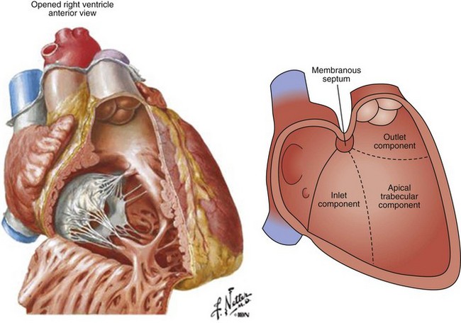
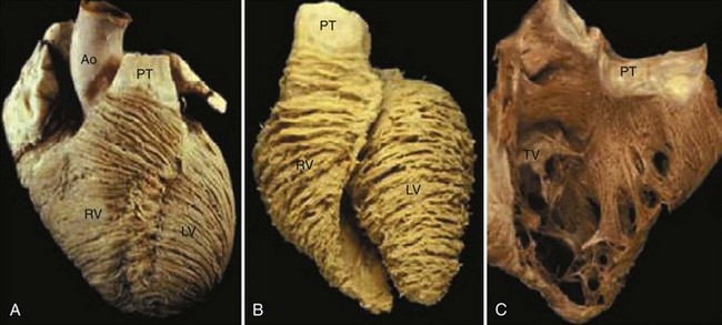
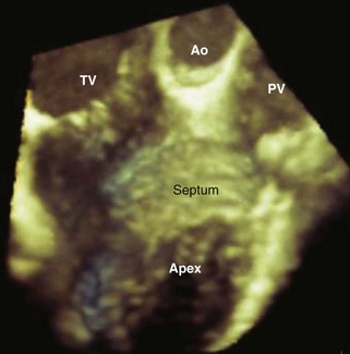
Figure 3-2 Anatomy of the RV, having three separate parts.
(Image on the left from Netter FH, Atlas of Human Anatomy, second edition. Philadelphia, Novartis, 1997; Plate 208.)
Coronary Artery
The coronary artery supply to the RV myocardium is shown in Figure 3-4, and the RV segments are shown in Figure 3-5.
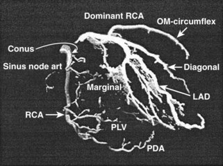
Figure 3-4 Coronary arteries to the RV.
(From Mangion JR. Right ventricular imaging by two-dimensional and three-dimensional echocardiography. Curr Opin Cardiol. 2010;22:423-429.)
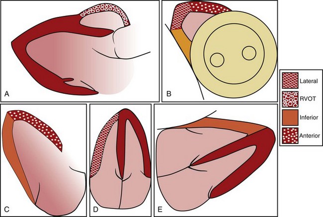
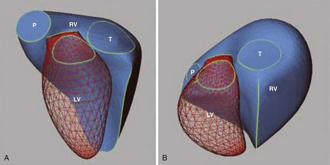
Figure 3-5 Segmentation of the RV wall.
(From Mangion JR. Right ventricular imaging by two-dimensional and three-dimensional echocardiography. Curr Opin Cardiol. 2010;22:423-429.)
Coronary flow to the RV is primarily from the right coronary artery (RCA) (dominant in 80% of the population) (Fig. 3-5):
Determinants of Right Heart Function
The function of the right heart is governed by the following four factors:
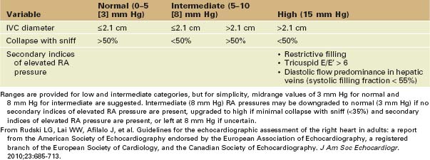
where RA pressure is estimated using the American Society of Echocardiography (ASE) standard (Table 3-1).
Importance of Assessing RV Function
RV size and function are important prognostic factors in many cardiopulmonary conditions as follows:
Echocardiographic Assessment of RV Size and Function
Stay updated, free articles. Join our Telegram channel

Full access? Get Clinical Tree


