18 Tuberculous Pleural Effusions
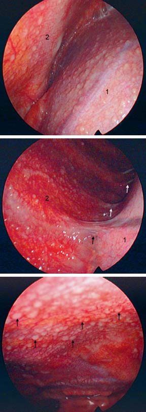
Fig. 18.1
a Tuberculous pleural effusion. After drainage of 800 mL of serous effusion: typical miliary sagolike nodules in all parts of the right parietal pleura, here on the diaphragm (1) and the anterior chest wall (2).
b Tuberculous pleural effusion. Same patient with firm adhesions (→) between the right lower lobe (1) and reddened, inflamed posterior chest wall (2), covered with sagolike miliary nodules.
c Tuberculous pleural effusion. Same patient with white miliary, sagolike nodules (→) on the anterior chest wall, which is highly inflamed.
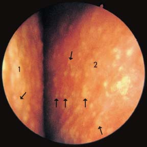
Fig. 18.2
Tuberculous pleural effusion. After drainage of 350 mL of serous effusion: small, miliary yellow-whitish nodules and patches of organized fibrin (→) on the pleural surface of the chest wall (1) and the lung (2).
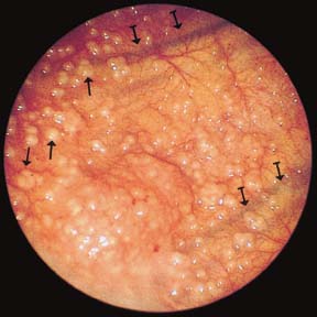
Fig. 18.3
Tuberculous pleural effusion. After drainage of 2500 mL of serous effusion: numerous small discrete nodules on all parts of the pleura. Here is seen a section of the chest wall with patchy granular surface consisting of nodules of different sizes (→). The vessels of the inflamed pleura are clearly dilated. Intercostal vessels and ribs ( ) show through the thickened parietal pleura.
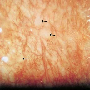
Fig. 18.4
Tuberculous pleural effusion. After drainage of 400 mL of opaque serous effusion, the visceral and parietal pleura show hyperemia and several fibrin patches. The photograph shows a magnification of a section of the chest wall, revealing serpiginous, partially distended, parallel vessels between which are relatively hyperemic, partially necrotic or fibrin-covered, millet-sized lesions (→).
Stay updated, free articles. Join our Telegram channel

Full access? Get Clinical Tree


