Tuberculosis and Nontuberculous Mycobacterial Infections

Chapter 31 Tuberculosis and Nontuberculous Mycobacterial Infections


The genus Mycobacterium consists of slow-growing organisms that are widely disseminated throughout the world and range from species that cause no human disease to those such as Mycobacterium tuberculosis and Mycobacterium leprae that are responsible for enormous morbidity and mortality. Mycobacteria are aerobic bacilli with high concentrations of lipids in their cell wall, which make them impermeable to most common stains. However, because of their ability to retain carbolfuchsin dye despite decolorization attempts with acid alcohol, they are referred to as acid-fast bacilli (AFB). Although mycobacteria can produce disease in almost any site, two groups of mycobacteria have a propensity for causing pulmonary infections: certain members of the M. tuberculosis complex and the nontuberculous mycobacteria (NTM).


Tuberculosis (TB) is the disease caused by bacteria of the M. tuberculosis complex, which includes the clinically relevant species M. tuberculosis, Mycobacterium bovis, and Mycobacterium africanum. Although M. tuberculosis is the most common cause of TB worldwide, both M. bovis and M. africanum can produce clinically indistinguishable forms of disease. The tubercle bacilli have been around for thousands of years, with evidence of human infection dating back to Neolithic, pre-Columbian, and early Egyptian times. Not until the Industrial Revolution, however, did TB become a major cause of human disease and death. It is estimated that approximately 25% of all adults died from TB in Europe during the 17th and 18th centuries. Throughout this period, the etiology of TB was hotly debated, with some of these early investigators arguing for a hereditary cause and others for a transmissible etiology. Finally, in 1882, Robert Koch presented his momentous discovery: The tubercle bacillus was the cause of TB. Early attempts at therapy, including the sanatorium movement, surgery, and lung collapse therapy, provided little relief from TB, and it was not until the discovery of paraaminosalicylic acid (PAS) and streptomycin in the 1940s that the age of antituberculosis chemotherapy began. Since that time, additional drugs have been developed, and most of the world treats TB with the same four-drug regimen administered for 6 months. More recently, however, co-infection with human immunodeficiency virus (HIV) and the emergence of drug-resistant strains of M. tuberculosis have conspired to complicate clinical management and to create barriers to global TB control.


The NTM group consists of nonlepromatous organisms that are not members of the M. tuberculosis complex. The NTM have been referred to as “mycobacteria other than tuberculosis” (MOTT), atypical mycobacteria, and environmental mycobacteria. The last designation refers to their widespread presence in the environment. The NTM have several features that distinguish them from M. tuberculosis: They have a wide range of pathogenicity, are not always associated with disease, and, unlike M. tuberculosis, are not transmissible from human to human. Of note, however, the incidence of NTM disease is increasing in many areas of the world, and the cause for this increase is unknown. Unfortunately, the pathogenic NTM are relatively drug resistant compared with M. tuberculosis, so NTM infections typically are difficult to treat. Because of the current poor understanding of the transmission and pathogenesis of these infections, little insight into their prevention has emerged, so no public health strategy for the control of disease caused by these ubiquitous organisms has been formulated.


Tuberculosis


Epidemiology, Risk Factors, and Pathogenesis


Epidemiology


The World Health Organization (WHO) estimates that 30% of adults worldwide are infected with organisms in the M. tuberculosis complex. From this large reservoir of infected people, an estimated 9 million new cases of TB occurred in 2009, leading to approximately 1.3 million deaths. In 2008, TB was estimated to be the seventh leading cause of death worldwide, and it is the number one killer of HIV-infected patients.


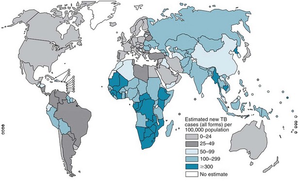
The burden of TB varies significantly throughout the world, with more than 90% of cases occurring among people residing in developing countries (Figure 31-1). The highest incidence rates for TB are in sub-Saharan Africa, particularly in the southern region of the continent. Not surprisingly, the highest prevalence of HIV co-infection also is in this region. Worldwide, approximately 23% of all persons with TB have underlying HIV co-infection; however, in sub-Saharan Africa, an estimated 50% of persons with TB have HIV/AIDS.


Recent reports of outbreaks of multidrug-resistant TB (MDR TB) and extensively resistant TB (XDR TB) have highlighted the importance of providing effective antituberculosis therapy to patients. MDR TB refers to disease caused by isolates of M. tuberculosis that are resistant to at least isoniazid (INH) and rifampin, whereas XDR TB refers to that due to MDR TB isolates that also are resistant to fluoroquinolones and at least one second-line injectable agent (amikacin, capreomycin, or kanamycin). Surveys have documented that approximately 4.6% of TB cases worldwide are MDR TB, and 5.4% of these cases are XDR TB. More cases of drug-resistant TB exist today than in all of recorded history, and this trend is likely to continue unless more effective TB control measures are implemented globally.


In the United States, the TB case rate declined by 3% to 5% per year from 1953 to 1984. Between 1986 and 1992, the numbers of TB cases increased by approximately 20%. This increase in the number of cases was the result of at least three major factors: (1) inadequate public health measures, (2) immigration from countries where TB is prevalent, and (3) co-infection with HIV. Fortunately, case numbers have declined since 1992 and are now at a historic low. In 2010, a total of 11,181 cases of TB were reported in the United States, for an incidence of 3.6 per 100,000 population. TB case rates declined an average of 4.5% each year during 2000 to 2010. TB rates were 11 times higher among foreign-born persons than among U.S.-born people. Among U.S.-born persons, blacks were 7 times more likely to have TB than whites. Approximately 1% of new cases in the country had MDR TB, approximately 90% of whom were foreign-born. Of these MDR TB cases, approximately 1% to 2% were XDR TB.


Risk Factors


Certain people are at higher risk for development of TB simply because they are more likely to be exposed to and thus infected with M. tuberculosis (Table 31-1). For example, approximately 60% of reported TB cases in the United States occur among foreign-born people who come from areas where the disease is endemic. Other populations with an increased prevalence of TB infection include certain racial and ethnic groups, low-income populations, the homeless, and injection drug users.


Table 31-1 Criteria for a Positive Reaction on Tuberculin Skin Testing


Induration Size Risk Groups
≥5 mm
≥10 mm
≥15 mm

* The predominant chest radiographic finding consistent with previous tuberculosis is presence of fibrotic lesions; other changes such as pleural thickening or isolated calcified granulomas are not related.


Medical conditions and factors associated with increased risk for development of active disease in a patient with latent tuberculosis infection include silicosis, end-stage renal disease, malnutrition, diabetes mellitus, carcinoma of the head or neck and lung, immunosuppressive therapy, lymphoma, leukemia, weight loss of more than 10% ideal body weight, gastrectomy, and jejunoileal bypass.


Anyone infected with M. tuberculosis can develop TB disease, but certain groups are at higher-than-normal risk for progression to active disease (see Table 31-1). Patients who have been recently infected with M. tuberculosis and those with medical conditions associated with significant immunosuppression are at particularly high risk for development of TB. HIV co-infection is the strongest known risk factor for the development of TB and is estimated to increase the risk of progression to TB by 50- to 100-fold. Inhibitors of tumor necrosis factor-α (TNF-α) may increase the risk for development of TB by up to 10-fold, and patients taking TNF-α blockers frequently present with disseminated disease. This association appears to be stronger for infliximab and adalimumab than for etanercept. Other medical conditions are associated with a more modest increase in risk for development of disease.


Pathogenesis


TB is spread from person to person almost exclusively through the air by droplet nuclei, which are particles 1 to 5 µm in diameter that contain viable tubercle bacilli. Droplet nuclei are expelled into the air when patients with infectious TB create an aerosol by talking, coughing, or singing. Three factors determine the likelihood of transmitting TB: the number of bacilli expelled into the air, the concentration of organisms in the air, and the duration of contact with (i.e., breathing of) the infected air. Whether an inhaled tubercle bacillus establishes an infection in the exposed person’s lung depends on both bacterial virulence and host immune defenses.


The tubercle bacillus grows slowly, dividing approximately every 18 to 24 hours. Tubercle bacilli spread through the lymphatics to the hilar lymph nodes or through the bloodstream. Small numbers of bacilli are deposited in other organs, which may then become sites of extrapulmonary disease. An adaptive immune response occurs after 2 to 8 weeks.


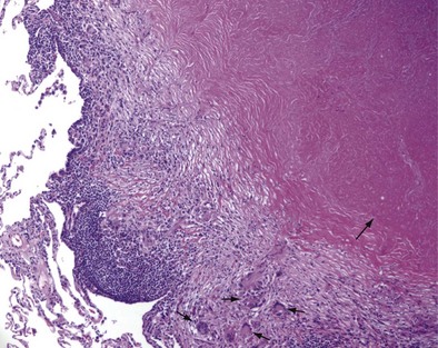
Once cell-mediated immunity develops, collections of activated T cells and macrophages form granulomas that wall off the mycobacterial organisms (Figure 31-2). For most persons with normal immune function, infection with M. tuberculosis seems to be arrested once cell-mediated immunity develops, even though small numbers of viable bacilli remain within the granuloma. Although a primary complex can sometimes be seen on chest radiograph, most TB infections are asymptomatic and can be detected only indirectly with a tuberculin skin test (TST) or interferon-γ release assay (IGRA). Persons with “walled-off” TB infection who do not have active disease are not infectious and thus cannot spread the disease to others.


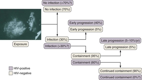
If cell-mediated immunity does not contain the tubercle bacilli, the initial infection progresses to active disease. Without treatment, infected persons have approximately a 5% chance of developing TB in the first 1 to 2 years after infection and an additional 5% chance of developing TB during the remainder of their lifetime (Figure 31-3). By contrast, persons who are co-infected with HIV have a 5% to 10% annual risk of active disease developing. When active TB develops soon after infection, the disease is referred to as primary TB. By contrast, when TB develops years or even decades after the initial infection, the disease is referred to as postprimary or reactivation disease. Exogenous reinfection, involving acquisition of a second strain of M. tuberculosis, also can lead to disease and seems to be more common in HIV-infected patients.


Clinical Features


The clinical manifestations of TB are protean. Patterns of disease vary depending on whether the disease is primary or reactivation in nature, the host’s immune status, and possibly the strain of M. tuberculosis. Of note, the clinical features of active TB are the result of a balance between host defenses and bacterial virulence; therefore, a continuum of disease is likely, and the clinical presentation of disease may be altered in severely immunocompromised patients. Most patients initially are seen with pulmonary disease, which is classically divided into primary disease and postprimary disease.


Pulmonary Tuberculosis


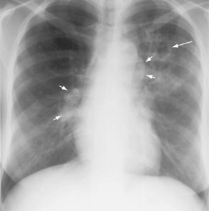
The initial infection in the lung, referred to as primary infection, causes formation of an inflammatory infiltrate, which may be seen on a chest radiograph, often in the middle or lower lung zones. The draining lymph nodes may enlarge and compress adjacent bronchi, particularly in infants and children. Parenchymal disease usually clears as cell-mediated immunity develops, and it tends to clear more rapidly than nodal involvement. If the parenchymal disease persists beyond the development of cell-mediated immunity, cavitation may occur, although this finding is uncommon. Pleural effusions are a common manifestation of primary TB and presumably result when a peripheral, caseous focus ruptures into the pleural space (Figure 31-4). Pleuritis caused by TB may manifest as an acute illness characterized by cough, fever, and pleuritic chest pain.


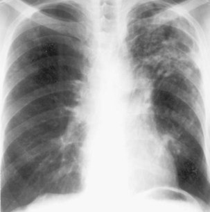
During most initial infections with M. tuberculosis, small numbers of organisms are disseminated hematogenously, and some become seeded in the apices of the lung. The organisms seem to grow preferentially in this well-oxygenated environment, with progression to active disease occurring months or years after the initial infection. This accounts for the characteristic radiographic location of reactivation disease, which in most cases occurs in the apical or posterior segments of the upper lobes (Figure 31-5). In areas of chronic infection or areas of caseation, fibrosis may occur. Fibrocaseous lesions may contain live mycobacteria for many years, and these are the lesions that may reactivate years later.


Pleural Tuberculosis


Pleurisy usually is a manifestation of primary TB and results when a subpleural caseous focus ruptures into the pleural space. The resulting delayed-type hypersensitivity reaction produces pleural liquid that has a high protein concentration. Most patients are initially seen with chest pain, fever, and a nonproductive cough. If left untreated, the pleural effusion will resolve spontaneously over 2 to 4 months. The rate of reactivation, however, is approximately 65% within the next 5 years.


Diagnosis of tuberculous pleural disease begins with sampling of the pleural fluid. Early in the course of disease, the fluid may have a polymorphonuclear predominance, but in almost all cases, mononuclear cells become the majority. Cell counts typically are in the 100 to 5000 cells/µL range, and the cells are almost all lymphocytes; the presence of mesothelial cells and/or eosinophils makes the diagnosis of TB unlikely.


Pleural fluid AFB smears are seldom positive, and pleural fluid cultures are positive in only approximately 20% to 40% of cases. M. tuberculosis can be isolated from 30% to 50% of induced sputum specimens from patients with TB pleuritis; such specimens should therefore be obtained in all patients. Pleural biopsy specimens provide the highest diagnostic yield, with positive culture results in up to 80% to 90% of cases when at least three specimens are obtained. Thoracoscopic biopsies are nearly always diagnostic, but the procedure is invasive, costly, and often not available.


Other tests that may be helpful in the diagnosis of pleuritis are adenosine deaminase and interferon-γ (IFN-γ) assays. The enzyme marker adenosine deaminase has been shown to have high sensitivity but variable specificity. A recent metaanalysis suggested that pleural fluid IFN-γ concentration has sensitivity of 89% and specificity of 97% for pleural TB in HIV-uninfected patients. Sensitivity and specificity also are high in HIV-infected patients.


When faced with a lymphocytic exudative pleural effusion in a patient with a positive TST reaction or IGRA result, the clinician should strongly consider TB. Whether to start treatment empirically or proceed with a pleural biopsy will depend on the certainty of the diagnosis and whether or not the patient is at risk for drug-resistant TB. In the latter situation, pleural tissue should be obtained for smear and culture to direct drug susceptibility testing.


Bone and Joint Tuberculosis


Skeletal involvement is thought to arise from reactivation from foci that were seeded at the time of initial infection. The infection begins in the subchondral region of the bone and then spreads to cartilage, synovium, and joint space. Although weight-bearing bones are the most likely to be affected, any bone or joint may be involved. In most series, TB of the spine, or Pott’s disease, accounts for more than 50% of cases. In children, the upper thoracic spine is the most frequently affected site, whereas in adults, the lower thoracic and upper lumbar vertebrae typically are involved. After the spine, the hips and knees are the most common sites of skeletal TB.


Most patients initially are seen with pain in the involved joint. Systemic symptoms usually are absent, and delays in diagnosis are common. Tuberculous involvement of the joint usually is first suspected after a radiograph shows changes suggestive of the diagnosis. Typical findings include metaphyseal erosion and cysts, loss of cartilage, and narrowing of the joint space. In Pott’s disease, two vertebral bodies and the intervening joint space usually are involved. CT and/or magnetic resonance imaging (MRI) should be obtained to better define the pattern and extent of involvement. Confirmation of the diagnosis requires aspiration of joint fluid or of periarticular abscesses or biopsy of affected bone or synovium. Acid-fast smears are positive in 20% to 25% of joint fluid aspirates, with isolation of mycobacteria in 60% to 80%. Histopathologic evidence of granulomatous inflammation is almost always present in bone and synovial biopsy specimens.


Diagnosis


To diagnose TB, the disease must first be suspected. TB should be suspected in certain high-risk groups reviewed previously (see Table 31-1) and when the clinical and/or radiographic presentation is consistent with TB. The medical history should elicit whether or not the person suspected of having TB has been exposed to M. tuberculosis or has a previous history of TB infection or disease. Symptoms at presentation will vary depending on the sites(s) of involvement and extent of disease as described previously. Guidelines suggest that all persons with an unexplained cough lasting 2 to 3 weeks or more be evaluated for TB. Of note, up to 20% of patients with pulmonary disease are asymptomatic. Findings at physical examination are rather nonspecific and will vary, depending on the site of involvement. Among HIV-infected patients, TB should be considered when any respiratory infection or fever of unknown origin occurs, because the risk for TB in this group is substantially elevated, and signs and symptoms of TB often are atypical.


Tuberculin Skin Test and Interferon-γ Release Assays


The TST (discussed in more detail later on), which uses purified protein derivative (PPD), is the most common way to identify persons with latent tuberculosis infection (LTBI) but should not be used in the diagnosis of active TB. In general, the sensitivity of the TST for detection of active TB ranges from 65% to 94%, but in critically ill patients with disseminated disease, the sensitivity decreases to only 50%. Thus, a negative TST reaction can never exclude a diagnosis of TB.


IGRAs measure the release of IFN-γ in whole blood in response to stimulation by M. tuberculosis antigens. Whole blood is incubated overnight with early secretory antigen target 6 [ESAT-6], culture filtrate protein 10 [CFP10], TB7.7, and control antigens; lymphocytes sensitized by previous exposure to M. tuberculosis release IFN-γ. IGRAs currently available include the QuantiFERON-TB (QFT-TB) Gold and QFT-TB Gold In-Tube (QFT-GIT), which measure IFN-γ in the serum using enzyme-linked immunosorbent assay (ELISA) (Cellestis Limited, Carnegie, Victoria, Australia) and the T-Spot.TB test, which uses enzyme-linked immunospot (ELISPOT) methodology (Oxford Immunotec, Oxford, United Kingdom) to identify IFN-γ–producing cells.


The reported sensitivity of QFT-GIT in patients with active TB has varied, ranging from 62% to 94%, with a pooled sensitivity of 80%, whereas the T-Spot.TB test has a sensitivity of 35% to 100%, with a pooled sensitivity of 81%. By contrast, the TST has a pooled sensitivity of 65% in patients with TB. Although the IGRAs have improved sensitivity compared with the TST, the values are still too low to rule out active TB with confidence, and neither test can differentiate latent from active TB.


Radiographic Examinations


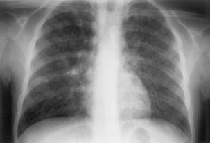
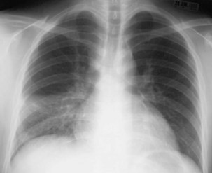
Plain chest radiography is a sensitive but nonspecific test to detect pulmonary TB. Radiographic manifestations of TB vary, depending on whether the patient has primary or postprimary TB and whether co-infection with HIV is present. Patients who have primary pulmonary TB at initial evaluation may demonstrate radiographic opacities in the lower lung zones and an associated pleural effusion (see Figure 31-4). TB caused by reactivation typically involves the apical and posterior segments of the upper lobes or superior segment of the lower lobe (see Figure 31-5). Cavitation and volume loss are common in reactivation disease but unusual in primary disease. Findings on the chest radiograph in patients co-infected with HIV depend on the severity of immunosuppression. Early in the course of HIV disease, the radiograph may show a typical reactivation pattern with cavitation (Figure 31-7), but as the CD4+ cell count declines, the radiographic appearance is more like the pattern seen in primary TB (Figure 31-8). Patients co-infected with HIV may sometimes have a normal-appearing chest radiograph despite being sputum AFB smear–positive.


Bacteriologic Examination


Sputum Microscopy


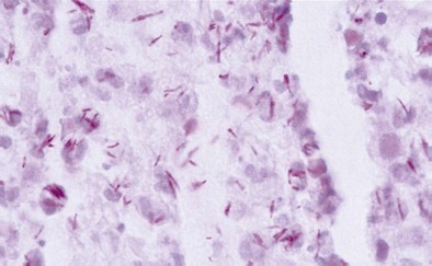
Diagnosis of pulmonary TB begins with obtaining two or three spontaneously expectorated sputum samples collected at 8- to 24-hour intervals, with at least one collected in early morning. Two methods are commonly used for acid-fast staining: the carbolfuchsin methods (Ziehl-Neelsen and Kinyoun methods) and a fluorochrome procedure that uses auramine O or auramine-rhodamine dyes (Figure 31-9).


Approximately 5,000 to 10,000 bacilli/mL are necessary to allow detection of these organisms in stained smears. The sensitivity of sputum AFB smears ranges from 50% to 80%, depending on the extent of disease; patients with cavitary disease are more likely than those without cavities to expectorate tubercle bacilli. Light-emitting diode (LED)–based fluorescence microscopy allows for more rapid evaluation of specimens and may increase the sensitivity slightly over that for conventional light microscopy. If patients are unable to produce sputum or have negative sputum smears, additional diagnostic tests may be indicated. In such circumstances, either sputum induction or biopsy using fiberoptic bronchoscopy (FOB) may provide adequate specimens. Studies suggest that sputum induction with hypertonic saline and FOB with bronchoalveolar lavage produce similar yields in smear-negative cases. The primary role of FOB is in smear-negative HIV-infected TB suspects, in whom this technique also can help identify alternative causes of the illness.


Jun 12, 2016 | Posted by in RESPIRATORY | Comments Off on Tuberculosis and Nontuberculous Mycobacterial Infections

Full access? Get Clinical Tree

Get Clinical Tree app for offline access