Pharmacologic Therapy
Antiarrhythmic drugs are not arrhythmia suppressants in the same way that menthol is a cough suppressant. They do not work by soothing irritable areas. In fact, most antiarrhythmic drugs work merely by changing the shape of the cardiac action potential. By changing the action potential, these drugs alter the conductivity and refractoriness of cardiac tissue. Thus, it is hoped, the drugs will change the critical electrophysiologic characteristics of reentrant circuits to make reentry less likely to occur.
Channels and Gates
Antiarrhythmic drugs are thought to affect the shape of the action potential by altering the channels that control ionic fluxes across the cardiac cell membrane. The class I antiarrhythmic drugs, which affect the rapid sodium channel, provide the clearest example (Figure 3.1).
Figure 3.1 The effect of class I drugs on the rapid sodium channel. (a) through (c) display the baseline (drug-free) state. In (a), the resting state, the m gate is closed and the h gate is open. The cell is stimulated in (b), causing the m gate to open, thus allowing positively charged sodium ions to enter the cell rapidly (large arrow). In (c), the h gate shuts and sodium transport stops (i.e. phase 0 ends). (d) and (e) display the effect of adding a class I antiarrhythmic drug (open circles). (d) shows the class I drug binding to the h gate, making it behave as if it were partially closed. When the cell is stimulated in (e), the m gate still opens normally, but the channel through which sodium ions enter the cell is narrower and sodium transport is slower. It subsequently takes longer to reach the end of phase 0 (f), and the slope of phase 0 is decreased.

The rapid sodium channel is controlled by two gates: the m gate and the h gate. In the resting state (a), the m gate is closed and the h gate is open. When an appropriate stimulus occurs, the m gate opens (b) and positively charged sodium ions rush into the cell rapidly, causing the cell to depolarize (phase 0 of the action potential). After a few milliseconds, the h gate slams shut (c), closing the sodium channel and ending phase 0.
Class I antiarrhythmic drugs work by binding to the h gate, making it behave as if it were partially closed (d). In this case, when the m gate is stimulated to open, the opening through which sodium ions enter the cell is narrower (e). Consequently, it takes longer to depolarize the cell (i.e. the slope of phase 0 is decreased). Because the speed of depolarization determines how quickly adjacent cells will depolarize (and therefore the speed of impulse propagation), class I drugs as a group tend to decrease the conduction velocity of cardiac tissue.
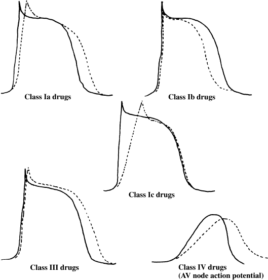
Although their precise sites of action have not all been worked out, most antiarrhythmic drugs act in a similar fashion: that is, by altering the function of the various channels that control transport of ions across cardiac cell membranes. The resultant changes in the cardiac action potential (Figure 3.2) cause changes in the conduction velocity, refractoriness, and automaticity of cardiac tissue, and also provide the basis for the classification of antiarrhythmic drugs.
Figure 3.2 The effect of antiarrhythmic drugs on the cardiac action potential. The solid lines represent the baseline action potential and the dotted lines represent the changes that result in the action potential when various classes of antiarrhythmic drug are given. The Purkinje fiber action potential is shown, except in the case of class IV drugs, for which the AV nodal action potential is depicted.

Classification of Antiarrhythmic Drugs
Table 3.1 lists the most frequently used classification system for antiarrhythmic drugs.
Table 3.1 Classification of antiarrhythmic drugs.
| Class I | Bind to sodium channel, decrease speed of depolarization | |
| Class II | β-blocking drugs, decrease sympathetic tone | |
| atenolol | nadolol | |
| bisoprolol | carvedilol | |
| labetolol | propranolol | |
| metoprolol | timolol | |
| Affect mainly SA and AV nodes (indirectly by blocking β receptors) | ||
| Class III | Increase action potential duration | |
| amiodarone | N-acetylprocainamide | |
| dronedarone | sotalol | |
| ibutilide | dofetilide | |
| Class IV | Calcium-channel blockers | |
| diltiazem | verapamil | |
| Affect mainly SA and AV nodes (direct membrane effect, see Figure 3.2) | ||
| Class V | Digitalis agents | |
| digitoxin | digoxin | |
| Affect mainly SA and AV nodes (indirectly by increasing vagal tone) | ||
Class I is reserved for drugs that block the rapid sodium channel (as shown in Figure 3.1). Because drugs assigned to class I block the sodium channel in varying degrees and also have varying effects on action-potential duration, class I is currently broken down into three subgroups (Table 3.2). Class Ia drugs (quinidine, procainamide, and disopyramide) slow conduction velocity and increase refractory periods. Class Ib drugs (lidocaine, tocainide, mexiletine, and phenytoin) actually have little effect on depolarization when used in systemic doses (although in high concentrations these drugs too can block sodium transport—this is why lidocaine is an excellent local anesthetic agent). In systemic doses, class Ib drugs decrease action-potential duration and shorten refractory periods but have little effect on conduction velocity. Class Ic drugs (flecainide, encainide, and propafenone) have a pronounced depressant effect on conduction velocity, with relatively little effect on refractory periods.
Table 3.2 Subclassification of class I antiarrhythmic agents.
| Class Ia | Quinidine, procainamide, disopyramide |
| Slow upstroke of action potential + + | |
| Prolong duration of action potential + + | |
| Decrease conductivity, increase refractoriness | |
| Class Ib | Lidocaine, phenytoin, tocainide, mexiletine |
| Minimal effect on upstroke of action potential | |
| Shorten duration of action potential | |
| Decrease refractoriness | |
| Class Ic | Flecainide, encainide, propafenone, moricizinea |
| Marked slowing of upstroke of action potential + + + + | |
| Minimal effect on action potential duration + | |
| Marked decrease in conductivity, little effect on refractoriness | |
| aThe classification of moricizine is controversial, and some place it in class Ib. It is placed in class Ic here to emphasize its class Ic-like proarrhythmic potential. | |
β-blocking agents are assigned to class II. These drugs have little direct effect on the action potential and work mainly by decreasing the sympathetic tone.
Class III drugs (amiodarone, dofetilide, dronedarone, ibutilide, N-acetylprocainamide, and sotalol) increase the action-potential duration and therefore refractory periods, and have relatively little effect on the conduction velocity.
Class IV includes the calcium-channel blockers. They mainly affect the SA and AV nodes, because these structures are almost exclusively depolarized by the slow calcium channels.
Class V includes digitalis agents, whose antiarrhythmic effects are related to the increase in parasympathetic activity caused by these drugs.
Effect of Antiarrhythmic Drugs
Most antiarrhythmic drugs are felt to ameliorate automatic tachyarrhythmias to some extent, although it must again be stressed that the primary treatment for automatic arrhythmias is to remove the underlying cause. Many antiarrhythmic drugs slow the phase 4 ionic fluxes that are responsible for automaticity.
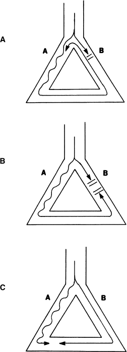
Figure 3.3 shows two examples of how antiarrhythmic drugs may affect reentrant circuits. Figure 3.3a shows a reentrant circuit with the same characteristics as described in Chapter 2. Figure 3.3b illustrates the changes that can occur if a class Ia drug is administered. These drugs increase refractory periods. By further lengthening the already long refractory period of pathway B, the class Ia drugs can convert unidirectional block to bidirectional block, thus chemically amputating one pathway of the reentrant circuit. Figure 3.3c shows what happens if a class Ib drug is given. These drugs shorten the duration of the action potential, thus decreasing the refractory period. In this example, a class Ib drug shortens the refractory period of pathway B, thus rendering the refractory periods in pathways A and B relatively equal. (In other words, the tachycardia zone is significantly narrowed.) Without a difference in refractory periods between the two pathways of the anatomic circuit, reentry cannot be initiated.
Figure 3.3 Effect of antiarrhythmic drugs on a reentrant circuit. (a) A prototype reentrant circuit (the same as that described in Figures 2.5 and 2.6). (b) Changes that may occur with administration of a class la drug. The refractory period of pathway B may be sufficiently prolonged by the drug to prevent reentry from occurring. (c) Changes that may occur with administration of a class lb drug. The refractory period of pathway B may be shortened, so that the refractory periods of pathways A and B are nearly equal. A premature impulse would then be more likely to either conduct or block down both pathways, thus preventing the initiation of reentry.

Stay updated, free articles. Join our Telegram channel

Full access? Get Clinical Tree


