Fig. 8.1
Type B 3 fracture and primary total elbow replacement

Fig. 8.2
Transcondylar humerus fracture (C2). Primary closed reduction and external fixator stabilization. Secondary open reduction and internal (anatomical) plate fixation after soft tissue consolidation
8.1.5 Elbow Luxations
The forearm most frequently dislocates posteriorly (posterolateral, posteromedial). Anterior dislocations are rare. The evaluation of the neurovascular status is mandatory. After reduction in analgo-sedation, the elbow should be immobilized (cast, >90°). The following treatment depends on the dynamic and static elbow stability. In case of biomechanical joint stability (coronoid intact), the therapy is nonoperative. Dynamic instabilities and coronoid fractures should be stabilized after soft tissue consolidation.
8.2 Proximal Forearm Fractures
8.2.1 Olecranon Fractures
Olecranon fractures are classified by Schatzker [9]. These fractures are tensile-loaded fractures; therefore tension band wiring is the standard procedure for simple fractures. For the stability it is very important to anchor the wires in the opposite corticalis. Proximal transverse fractures may be fixed by cancellous compression screws. Plate fixation is used for comminuted fractures (LCDCP/LCP).
8.2.2 Radial Head Fractures
The treatment is based on the Mason classification. Type 1 fractures are treated nonoperatively. Type 2 fractures are indication for open reduction and internal mini-screw fixation. Closed reduction and titanium elastic nail fixation may be performed in displaced fractures of the radial neck (Metaizeau technique) [10]. Open reduction and mini-plate fixation are used in type 3 fractures. Radial head resection is an option in comminuted factures. The radial head replacement is necessary in case of instability of the lateral column to prevent a proximal migration of the radius and in case of laceration of the interosseous membrane (Essex-Lopresti fracture).
8.2.3 Forearm Shaft Fractures
Ulna and radial parry fractures are distinguished from forearm fractures. In general nonoperative therapy is possible. However, surgery offers the opportunity of functional treatment. The classic forearm fracture is an absolutely indication for surgery. A2 and A3 fractures have to be fixed under compression with LCDC plates to avoid pseudarthroses. Locking plate systems are used to stabilize A1, B, and C fractures. The stabilization starts with the ulna fracture by using a medial, intermuscular approach. Thompson’s approach is the standard approach for radius shaft fractures [11]. In case of very proximal radius fractures, the Henry approach is recommended [12]. It is very important to protect the profund branch of the radial nerve. Unimpeded pro- and supination should be documented after plate fixation.
8.2.4 Monteggia Fractures
Fracture of the proximal third of the ulna with dislocation of the radial head.
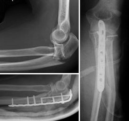
Bado describes four types, depending on the dislocation of the radial head. The Monteggia-like lesion describes an additional radial head fracture. Usually, the anatomical reduction of the ulna fracture leads to a spontaneous radial head reduction. LC and LCDC plates are used for internal fixation. The humeroradial joint should be explored in case of instability, and the annular ligament should be reconstructed (Fig. 8.3).


Fig. 8.3
Monteggia-like lesion. Open reduction and plate fixation of the ulna. Mini-screw fixation of the radial head
8.2.5 Galeazzi fractures
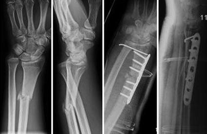
Fracture of the radius shaft with dislocation of the ulnar head. Open reduction and internal plate fixation (LCDCP) of the radius lead to the reduction of the distal radioulnar joint (DRUJ). In case of DRUJ instability, temporary transfixation may be necessary (K-wires and long arm cast for 8 weeks) (Fig. 8.4).


Fig. 8.4
Galeazzi fracture. Open reduction and internal radius plate fixation. K-wire transfixation of the DRUJ
8.2.6 Essex-Lopresti Fractures
The Essex-Lopresti lesion represents a complex distorsion injury of the wrist, comparable to the mechanism of a Maisonneuve fracture. The Essex-Lopresti fracture is a combination of DRUJ dislocation, laceration of the interosseous membrane, and radial head fracture [13]. These unstable injuries need surgery: temporary transfixation of the distal radioulnar joint, internal fixation of the radial head, and in case of comminuted fractures a radial head replacement.
8.2.7 Distal Radial Fractures
Distal radius fractures are the most common fractures (25 %), frequently seen in osteoporotic patients. Depending of the position of the hand during a fall, different fracture types are distinguished. The aim of the treatment is to restore the 3 columns of the wrist anatomically (reconstruction of the Böhler’s angle). Palmar locking plate fixation is recommended for type A2, A3, B2, B3, C1, and 2.3 fractures. Chauffeur fractures (B1) are fixed by compression screws. The intrafocal (Kapandji) or extrafocal (Willenegger) K-wire osteosynthesis may be used in A2 fractures or in critical soft tissue conditions, when open reduction is not possible. K-wires are also used in addition to plates or external fixators in case of destruction of the articulating surface. Bridging external fixators are useful in osteoporotic-comminuted fractures, precarious soft tissue conditions, high-grade open fractures, and polytraumatized patients. Additional injuries like SL—dissociations, displaced fractures of the base of the ulna styloid, and TFCC—and DRUJ instabilities require additional surgery.
8.3 Lower Limb Injuries
8.3.1 Femoral Head Fractures
Fractures of the femoral head (frequently combined with dorsal hip dislocation or femoral neck fractures) are the result of a high-energy trauma (e.g., dash board injury). The Pipkin classification describes 4 types [14]. The indication for open reduction and internal screw fixation of type 1 fractures depends on the extent of head destruction, fragment displacement, and impingement. Surgery is indicated in all type 2–4 cases. The main complications resulting from femoral head fractures are AVN of the femoral head, heterotopic ossification, and secondary coxarthrosis.
8.3.2 Femoral Neck Fractures
In the times of conservative treatment, femoral neck fractures had an extremely high mortality rate due to immobilization-related complications (DVT, pulmonary embolism, pneumonia).
Even today femoral neck fractures of the elderly show 1-year mortality of up to 25 %.
The Pauwels classification, defining the degree of instability based on the angle between the fracture and the horizontal plane, was used to identify stable fractures, which could be treated conservatively, and unstable fractures, which needed surgery, have become obsolete, as most fractures today are treated with primary hip replacement. The Garden classification considers the extent of the displacement and is used to estimate the prognosis of the fracture regarding the risk of nonunion and AVN.
Because of the compromised blood supply of the femoral head, only minimal displaced neck fractures in younger patients stand a chance of unimpaired fracture healing. These fractures should be reduced and fixed with cannulated screws preferably within the first 6 h to ensure best results. Operative decompression of the hip joint is controversial.
Hip replacement is recommended within 24–36 h. Studies did not show any significant differences between the use of bipolar prostheses or total hip replacement [15].
8.3.3 Trochanteric Fractures
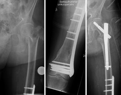
According to the AO classification, trochanteric fractures are classified into stable and unstable fractures. There are three types of instabilities: mediolateral, craniocaudal, and rotational instability. The posteromedial support (lesser trochanter defect) and the involvement of the subtrochanteric region are crucial for the stability. The mini-open reduction and extramedullary stabilization (e.g., by DHS) may be used for stable trochanteric fractures (31A1). Intramedullary nailing with cephalomedullary nails has become the gold standard for all types of intertrochanteric and subtrochanteric fractures, as these nails are suitable for both stable and unstable fractures.
Fixation of the lesser trochanter is not necessary. In displaced fractures with involvement of the subtrochanteric region, open reduction and additional internal cable wire fixation will increase the stability (Fig. 8.5).


