CHAPTER 5 Transoesophageal and stress echo and other echo techniques
5.1 TRANSOESOPHAGEAL ECHO
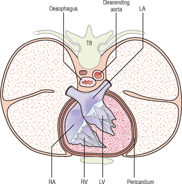
The echo techniques described so far have used ultrasound directed from the chest wall – transthoracic echo (TTE). The oesophagus in its mid-course lies posterior to and very close to the heart and ascending aorta and anterior to the descending aorta (Fig. 5.1).
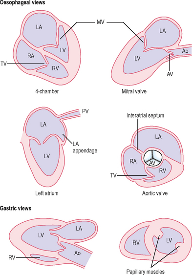
An echo technique exists for examining the heart with a transducer in the oesophagus – transoesophageal echo (TOE) (Figs 5.2, 5.3, 5.4, 5.5). In some countries, the abbreviation used is TEE. This uses a transducer mounted upon a modified probe similar to those used for upper gastrointestinal endoscopy and allows examination of the heart without the barrier to ultrasound usually provided by the ribs, chest wall and lungs. By advancing the probe tip to various depths in the oesophagus and stomach, manoeuvring the tip of the transducer and by altering the angle of the ultrasound beam with controls placed on the handle, a number of different views of the heart can be obtained.

Fig. 5.4 Structures at aortic valve level on short-axis view using TOE. The interatrial septum is shown (arrow).

Fig. 5.5 Short-axis view of left ventricle using TOE – transgastric view. AW, anterior wall; C, cavity; IVS, interventricular septum; LVPW, left ventricular posterior wall; LW, lateral wall.
Advantages of TOE
Disadvantages of TOE
Because of the invasive nature of TOE, it should only be performed if there is a good indication and after TTE has been performed. TOE-derived information should be used to complement that derived from TTE and not as an alternative. The potential risks of TOE (e.g. oesophageal damage) should be weighed up carefully against the potential benefits.
Uses of TOE
Patient preparation and care during TOE
The patient should give informed consent being aware of the potential risks which include:
The patient should have fasted for at least 4 h. All false and loose teeth should be removed. There should be no history of difficulty in swallowing solids or liquids (dysphagia) which might suggest oesophageal disease. It is advisable to give oxygen during the procedure via nasal cannulae, to monitor blood oxygen with a pulse oximeter and to have suction equipment available to remove saliva from the mouth. Continuous ECG monitoring should be carried out as with any echo examination. Resuscitation equipment should be available.
A local anaesthetic spray (e.g. lidocaine (lignocaine) 10%) is used on the pharynx. Several sprays are given and there may be some systemic absorption. Intravenous sedation with a short-acting agent such as the benzodiazepine midazolam is often used. The patient is placed in the left lateral position with the neck fully flexed to aid insertion of the transducer into the oesophagus. A plastic bite guard is placed in the mouth to protect the transducer and the fingers of the person performing the TOE.
It is unusual to need to give a general anaesthetic (e.g. if TOE is considered essential and the patient is unable to tolerate the procedure under local anaesthesia and i.v. sedation). TOE is often carried out as a day-case procedure. After the procedure, the patient should not eat or drink for at least 1 hour (to prevent aspiration into the lungs or burning of the throat) since the throat remains numb and the patient may still be drowsy.
Contraindications to TOE
Specific uses of TOE
1. Cardiac or aortic source of embolism
TOE is often carried out in young patients (age <50 years) who have had a stroke. Approximately 20% may have a cardiac embolic source.
Detection of intracardiac thrombus with TTE is difficult with a high false-negative rate despite high suspicion on clinical grounds. TOE is superior not only because of improved image resolution but also because it is better at viewing areas where thrombus is likely to occur, such as LA appendage. This is the commonest site for thrombus, usually in patients with underlying heart disease.
Risk factors for LA thrombus include:
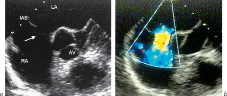
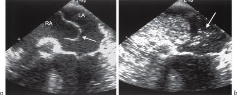
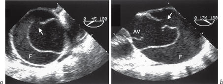
In some studies of patients with cerebral ischaemia (TIA and stroke), up to 5% had LA thrombus and in 75% of cases this was in the LA appendage (Fig. 5.6). Thrombus may appear as a rounded or ovoid mass that may completely fill the appendage. False-positive diagnosis of thrombus may occur due to misinterpretation of LA anatomy:

Fig. 5.6 Thrombus in left atrial appendage (arrow). There is also spontaneous echo contrast in the left atrial cavity.
Spontaneous echo contrast
A swirling ‘smoke-like’ pattern of echo densities within any cardiac chamber is known as spontaneous contrast. It is usually seen in low-output states. It is most often seen in the LA in mitral disease (up to one-third of cases), especially MS where it may occur in up to 50% of cases. It is due to sluggish flow and is associated with clumping of red cells (rouleaux formation) which become more echo-reflective. There is an increased thromboembolic risk – LA thrombus occurs in 20–30% of those with spontaneous contrast.
Other LA structural abnormalities associated with increased thromboembolic risk include ASD, patent foramen ovale (PFO) and atrial septal aneurysm.
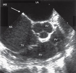
Atrial septal aneurysm (Fig. 5.7)
This is a bulging of the fossa ovalis and is found at autopsy in 1% of individuals. For echo purposes, the bulge must involve 1.5 cm of the septum and protrude 1.1 cm into either atrium. It is found in 0.2% of TTE series. In suspected cardiac source of embolism, it occurs in up to 15% of cases. The association with TIA/stroke may be because the aneurysm is thrombogenic and/or due to its frequent association with PFO and ASDs, which may allow paradoxical right to left embolization. TOE can help to detect all of these. A bubble contrast study during TOE can help to identify a small ASD or PFO and show a small shunt (section 6.4).

Fig. 5.7 Atrial septal aneurysm on TOE study. (a) Aneurysm bulging into left atrium (arrow). There appears to be a defect at lower region of aneurysm, probably a patent foramen ovale. (b) Bubble contrast study showing bubbles crossing from right to left atrium (arrow) through an associated patent foramen ovale.
TOE can show thrombus in other parts of the heart, e.g. LV mural thrombus. This is detected in over 40% of cases of acute MI at autopsy. Usually this occurs in the presence of anterior infarction and apical dyskinesis or LV aneurysm. Thrombus can also form in other low-output states, especially with chamber enlargement or where there is foreign material in the heart, e.g. pacing leads, central lines, prosthetic valves, particularly if inadequately anticoagulated or malfunctioning.
2. Examination of the aorta
TTE only gives good images of the ascending aorta, aortic arch and proximal descending aorta in a small minority of adults. TOE can add to this by providing excellent imaging of the aortic root, proximal ascending aorta, distal aortic arch and descending thoracic aorta. The interposition of the trachea between the oesophagus and ascending aorta limits the ability to image the upper ascending aorta and proximal aortic arch.
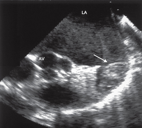
Aortic dimensions and dilatation
TOE allows accurate determination of aortic dimensions and reveals dilatation seen in aortic aneurysm.
Aortic atheroma
TOE helps detect and differentiate mobile and immobile atheromatous plaques. Mobile plaques may be associated with a higher embolic rate, as are pedunculated rather than linear plaques. Atheromatous plaques in the ascending aorta are found by TOE in at least 1% of individuals who have suffered an embolic CVA.
3. Endocarditis
TTE should always be used in the initial assessment of suspected or definite endocarditis. The superior spatial resolution provided by TOE allows small vegetations of only 1–2 mm to be identified and their location and morphology to be examined. All valves can be examined, but TOE is especially useful for the mitral and aortic valves (right-sided vegetations are often large and can be detected by TTE). In aortic subacute bacterial endocarditis (SBE), TOE is especially useful for aortic root abscess (TOE shows over 85% of such cases, TTE less than 30%), fistula or aneurysm of the sinus of Valsalva.
TOE is of use in endocarditis:
TOE should be considered in the majority of cases of suspected endocarditis.
4. Native valve assessment
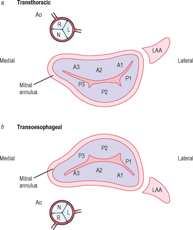
Mitral valve
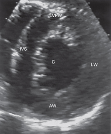
TTE is good but some aspects may be hard to assess. The posterior leaflet may be poorly visualized, especially if calcified or in the presence of mitral annular calcification. TOE can provide essential information in planning intervention such as MV repair (Figs 5.9 and 5.10).

Fig. 5.9 TOE showing (a) severe prolapse of posterior mitral valve leaflet (arrow) and (b) severe mitral regurgitation.

Fig. 5.10 Mitral valve viewed from (a) transthoracic parasternal view and (b) transoesophageal mid-gastric view. The 3 scallops of the anterior (A1, A2, A3) and posterior (P1, P2, P3) leaflets, the left atrial appendage (LAA) and the position of the proximal aorta (Ao) are shown. N = non-coronary sinus, R = right coronary sinus, L = left coronary sinus.
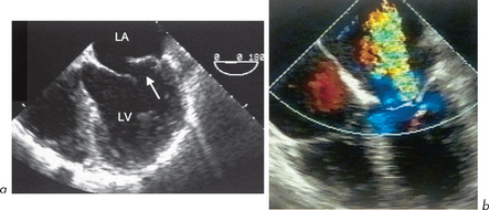
In MR, quantitative assessment of severity by TTE is difficult. TOE allows a more thorough assessment by Doppler and colour flow of the degree of MR within the LA. Severity can also be assessed by the pattern of pulmonary venous flow (severe MR may be associated with reversal of flow). The morphology of the valve can be examined to assess if suitable for valve repair rather than replacement. The exact segment of the valve which is causing regurgitation can be identified.
TOE can be used intraoperatively to assess the adequacy of valve repair.
In MS, TOE is very useful in deciding if a stenosed mitral valve is suitable for balloon valvuloplasty or whether surgical treatment such as mitral valvotomy or replacement is needed.
Balloon valvuloplasty for MS is not suitable if:
Aortic valve
TOE allows confident prediction of the integrity and number of cusps, evaluation of the aortic root, aortic sinuses and LVOT. Morphological assessment of AV can help give an indication of the aetiology of AR and colour flow mapping gives an indication of severity.
Tricuspid and pulmonary valves and right heart
The TV does not lend itself particularly well to TOE. Views can be obtained, but TTE is often sufficient. The PV, right ventricular outflow tract (RVOT) and proximal pulmonary artery can also be imaged reasonably well by TOE. It is often possible to view the 4 pulmonary veins and their connections with the LA, or to determine if there is partial or total anomalous pulmonary venous drainage.
5. Prosthetic valve assessment (see section 6.3)
The MV position is particularly well examined because of the orientation relative to the transducer. Paravalvular MR is well detected and may occur in up to 2.5% of all MV prostheses. TOE can be used intraoperatively and postoperatively to assess the presence and severity of paraprosthetic MR. TOE is useful in distinguishing between mild, moderate and severe paraprosthetic MR (the latter may deteriorate progressively and require re-operation). Shadowing of the LVOT occurs with mitral prostheses and may limit the ability to detect AR.
For aortic prostheses, TOE also has advantages over TTE, especially in biological valve degeneration, obstruction of prosthesis, regurgitation, abscesses or mass lesions (vegetations, thrombus). There are still some limitations even with TOE. The imaging planes are limited and as a result the acoustic shadow generated by mechanical prostheses may hide lesions in some areas. Aortic prostheses leave a portion of the aortic annulus immune from interrogation which may lead to underdiagnosis of root abscess.

Full access? Get Clinical Tree

