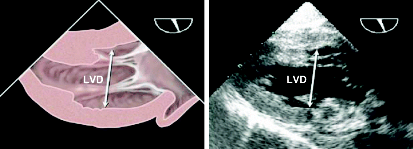
Figure 31 Transesophageal echocardiographic measurements of left ventricular (LV) minor-axis diameter (LVD) from transgastricric 2-chambers view of LV, usually best imaged at angle of approximately 90 to 110 degrees after optimizing maximum obtainable LV size by adjustment of medial-lateral rotation. (Reproduced, with permission, from Lang et al., 2005)


