Transcatheter Closure of Atrial Septal Defects and Patent Foramen Ovale

20 Transcatheter Closure of Atrial Septal Defects and Patent Foramen Ovale


An atrial septal defect (ASD) is a communication between the right atrium and left atrium due to an abnormal septation. There are four types of ASD:


ASDs are among the most common congenital heart defects. At present, only the secundum type is amenable to transcatheter closure.


Secundum ASD


This is a defect or deficiency in the septum primum in which the overlap with the septum secundum is incomplete, leading to the defect. The defect is therefore bordered by the limbus of the fossa ovalis or the C-shaped septum secundum. It comprises 10% of all congenital heart defects and is twice as common in females as in males. Associated anatomical lesions include mitral valve prolapse, partial anomalous pulmonary venous return, and complex congenital heart defects.


The hemodynamic pathophysiology typically involves a left-to-right shunt at the atrial level. The flow across an ASD or other atrial level shunt occurs mainly in diastole, and its direction depends on the differences in the atrial pressures and compliance rather than on pulmonary vascular resistance (PVR) or systemic vascular resistance (SVR), although these resistances are indirect factors. The compliance of the atria is determined by their respective ventricular compliance, which is dependent on ventricular wall thickness, a function of ventricular pressure and the resistance (e.g., PVR for the right ventricle and SVR for the left ventricle). As right ventricular pressure and PVR increase, the wall thickness of the right ventricle increases, leading to a fall in right ventricular and right atrial compliance (i.e., atrial wall becomes stiffer). Normally the mean left atrial (LA) pressure is 6 to 9 mmHg and the mean right atrial (RA) pressure is 1 to 4 mmHg. This favors a left-to-right shunt.


The pressure in the respective atria is dependent on the compliance of the respective ventricles. If the ventricles are poorly compliant or stiff, a higher atrial pressure is required for filling to occur. Unless the communicating defect is small, the amount of flow is dependent on the difference in compliance of the ventricles rather than the pressure, since a large defect will equalize the pressures in the atria. Normally, the right ventricular compliance is much higher than that in the left ventricle, resulting in a left-to-right shunt across the ASD, and its magnitude is dependent on the relative difference between the right ventricular and left ventricular compliance.


Left ventricular compliance is relatively stable for the first 20 to 30 years of life. As aging occurs, the arteriolar elasticity decreases and SVR increases, leading to higher blood pressure. This leads to higher energy expenditure by the left ventricle, to overcome increased afterload, and subsequent left ventricular hypertrophy. The compliance of the left ventricle decreases, with a subsequent elevation in LA pressure and increased left-to-right atrial level shunt. The shunt results in right-sided volume overload. Thus, the right atrium, right ventricle, pulmonary arteries, and pulmonary vascular bed are enlarged because of the increased volume of the shunt. There is increased flow across an otherwise normal tricuspid and pulmonary valve, leading to increased turbulence or a flow-related gradient across these valves.


Clinical Presentation


Most children with an ASD present with a murmur and are asymptomatic. Occasionally, infants may present with breathlessness, recurrent chest infections, and even heart failure. Failure to thrive is an uncommon presentation.


Adults with an ASD typically have a prolonged asymptomatic course. Symptom onset is insidious, most often occurring after the age of 40 or 50. Women may become symptomatic during the physiologic demands of pregnancy or labor. In adults with an ASD who are less than 40 years of age, there is no correlation between symptoms (New York Heart Association [NYHA] class) and the size of a shunt. Even patients with small (<10 mm) defects can present with significant symptoms. However, the development of symptoms does correlate with age. Major and limiting problems are often experienced after age 65 years.


The clinical course of an unrepaired ASD in adulthood may be significantly affected by hypertension, coronary artery disease, and mitral regurgitation. Patients with unrepaired ASD over 60 years of age often develop atrial fibrillation, an age-related reflection of atrial stretch, which seldom occurs in those younger than 40 years of age.


Symptoms may include the following:


The physical examination findings depend on the stage of presentation and pathophysiology. For example, cyanosis suggests severe pulmonary hypertension with reversed shunting in the presence of a secundum ASD or superior sinus venosus defect. A diastolic murmur at the lower right sternal border suggests increased blood flow through the tricuspid orifice, especially if the Qp:Qs ratio is more than 2.5:1 (relative tricuspid stenosis). The clinical findings and auscultation may also be completely unremarkable.


Echocardiography


In the current era of percutaneous device closure of interatrial communications, evaluation with transesophageal echocardiography (TEE) or intracardiac echocardiography (ICE) is mandatory prior to consideration of device closure.


Initial screening imaging with transthoracic echocardiography (TTE) may demonstrate a clearly visible defect in the atrial septum, best seen in the apical four chamber and subcostal long-axis views. However, it is common to see “echo dropout” in the region of the interatrial septum, and this may lead to misdiagnosis. Bubble contrast echocardiography with provocation (e.g., including a sharp nasal sniff, a cough, or the relaxation phase of the Valsalva maneuver) improves diagnostic accuracy. A positive test reveals the rapid transit of bubbles from the right to the left heart within three to five cardiac cycles. The amount of bubbles seen is related to the size of the defect. Late transit (more than five cardiac cycles) of bubbles is associated with intrapulmonary shunting. Intravenous contrast injection in the left arm may diagnose a persistent left SVC with anomalous drainage to the left atrium.


Table 20-1 summarizes the key points regarding TEE evaluation of intracardiac anatomy.


Table 20-1 Transesophageal Echocardiogram Features to Consider for Atrial Septal Defect Closure


Cardiac catheterization is not usually required for diagnosis. However, in older patients a diagnostic cardiac catheterization with full hemodynamic and coronary assessment may be justified.


Indications for Percutaneous Closure of Secundum ASD


Large ASDs should be closed to reduce the risk of complications; these may include premature death, atrial arrhythmias, reduced exercise tolerance, hemodynamically significant regurgitation, right-to-left shunting and embolism during pregnancy, congestive heart failure, or pulmonary vascular disease. Large defects with evidence of right ventricular volume overload on echocardiography may only cause symptoms in the third decade of life or beyond; however, regardless of symptoms, closure is usually indicated to prevent long-term complications.


Symptoms or complications of an ASD are indications for closure regardless of age. ASD closure will prevent further deterioration and probably will reverse or normalize right ventricular dilation right ventricular failure, and tricuspid regurgitation (TR). Atrial flutter or fibrillation after defect closure may be treated with radiofrequency ablation, ablation of the cavotricuspid isthmus, or atrial surgery (Maze procedure).


If the patient presents with pulmonary arterial hypertension (PAH), complete assessment of the reversibility of pulmonary vascular disease should be done prior to closure. Closure may be considered in the presence of net left-to-right shunting, pulmonary artery pressure less than two-thirds systemic levels, PVR less than two-thirds SVR, or when PAH is responsive to either vasodilator therapy or test occlusion of the defect. Patients should be treated in conjunction with providers who have expertise in the management of pulmonary hypertensive syndromes.


Closure of an ASD also is reasonable in the presence of paradoxical embolism and documented orthodeoxia-platypnea.


Closure of ASD should be considered in some cases as prophylaxis even if the defect is small. For example, patients who are professional divers or patients undergoing pacemaker implantation will benefit due to a reduced risk of paradoxical embolism.


Pregnancy and delivery are generally well tolerated, even by patients with an unclosed ASD with a significant left-to-right shunt. However, clinical symptoms may emerge during pregnancy or after childbirth. During pregnancy and delivery there is an increased risk of paradoxical embolism, regardless of the defect size. In our practice we close the defect before planned pregnancy, even if it is hemodynamically insignificant.


Contraindications for Percutaneous Closure of Secundum ASD


Small ASDs with a diameter of <5 mm and no evidence of right ventricular volume overload do not impact the natural history of the individual and do not require closure unless associated with paradoxical embolism.


An absolute contraindication for percutaneous ASD closure is the presence of severe and irreversible PAH, with no evidence of a left-to-right shunt. See Table 20-2 for a summary of the indications and contraindications to ASD closure.


Table 20-2 Indications and Contraindications to Atrial Septal Defect Closure


Indications

Contraindications


ASD Closure Devices


Currently the only two devices approved for the percutaneous closure of secundum ASD are the Amplatzer septal occluder and HELEX septal occluder device.


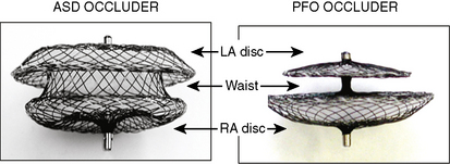
Amplatzer Septal Occluder (ASO)


The Amplatzer septal occluder (ASO) is a self-expandable double disc device made of nitinol (55% nickel, 45% titanium) wire mesh. The ASO is tightly woven into two flat discs (Fig. 20-1). There is a 3- to 4-mm connecting waist between the two discs, corresponding to the thickness of the atrial septum. Nitinol has super elastic properties, with shape memory. This allows the device to be stretched into an almost linear configuration and placed inside a small sheath for delivery and then return to its original configuration within the heart when not constrained by the sheath. The device size is determined by the diameter of its waist and is constructed in various sizes ranging from 4 to 40 mm (1-mm increments up to 20 mm; 2-mm increments up to the largest device currently available, 40 mm; the 40-mm size is not available in the United States). The two flat discs extend radially beyond the central waist to provide secure anchorage.


The LA disc is larger than the RA disc. For devices 4 to 10 mm in size, the LA disc is 12 mm and the RA disc is 8 mm larger than the waist. However, for devices larger than 11 mm and up to 32 mm in size, the LA disc is 14 mm and the RA disc is 10 mm larger than the connecting waist. For devices >32 mm, the LA disc is 16 mm larger than the waist and the RA disc is 10 mm larger than the waist. Both discs are angled slightly toward each other to ensure firm contact of the discs to the atrial septum.


A total of three Dacron polyester patches are sewn securely with polyester thread into each disc and the connecting waist to increase the thrombogenicity and endothelialization of the device. A stainless steel sleeve with a female thread is laser-welded to the RA disc. This sleeve is used to screw the delivery cable to the device. Each device costs US $5500.


Step-by-Step Technique: Transcatheter Device Closure of Secundum ASD


Method


1. Preprocedure. Review all pertinent data relating to the patient and to the defect to be closed and ensure that appropriate devices and delivery systems are available. The procedure and complications should be explained and opportunity given to ask questions. All preprocedure orders should be given to the patient. Aspirin 81 to 325 mg should be started 48 hours prior to the procedure. If allergic to aspirin, clopidogrel 75 mg should be used.


2. Vascular Access. The right femoral vein is accessed using a 7F or 8F short sheath. An arterial monitoring line (e.g., 4F) can be inserted in the right femoral artery, especially if the patient’s condition is marginal or if the procedure is performed under TEE and general endotracheal anesthesia. If a subclavian or internal jugular venous approach is used, it is very difficult to maneuver the device deployment, especially with large defects.


Heparin IV (e.g., 40U/kg) is given to achieve an activated clotting time (ACT) of more than 200 seconds at the time of device deployment. Antibiotic coverage for the procedure is recommended (e.g., cefazolin 1 g IV), the first dose at the time of procedure and two subsequent doses 6 to 8 hours apart.


3. Routine right heart catheterization should be performed in all cases to ensure presence of normal PVR. The left-to-right shunt can also be calculated.


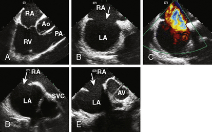
4. Echocardiographic assessment of the secundum ASD is performed simultaneously using either TEE or ICE. Figure 20-2 demonstrates full assessment of the defect by ICE.


The important ASD rims to look for are:


The rims must be sufficient (>5 mm) except for the anterior rim. A deficient anterior rim is not a contraindication to the procedure.


5. How to Cross the ASD. Use a multipurpose catheter. The MP A2 catheter has the ideal angle. Place the catheter at the junction of the IVC and the right atrium. The IVC angle should guide the catheter to the ASD. Keep a clockwise torque on the catheter while advancing it toward the septum (posterior). If unsuccessful, place the catheter in the SVC and slowly pull it into the right atrium; keep a clockwise posterior torque to orient the catheter along the atrial septum until it crosses the defect. TEE/ICE can be very useful to guide the catheter across difficult defects.


6. Perform a right upper pulmonary vein angiogram (Fig. 20-3A) in the hepatoclavicular projection (35-degree left anterior oblique/35-degree cranial). This delineates the anatomy, shape, and length of the septum. This may come in handy when the device is deployed but not released—the operator can position the imaging tube in the same view of the angiogram and compare the position of the device with that obtained during the deployment (Fig. 20-3B, C).


Jun 5, 2016 | Posted by in CARDIAC SURGERY | Comments Off on Transcatheter Closure of Atrial Septal Defects and Patent Foramen Ovale

Full access? Get Clinical Tree

Get Clinical Tree app for offline access