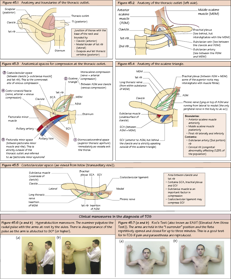
Thoracic outlet syndrome (TOS) is a neurovascular compression at the level of the thoracic outlet.
Aetiology
There are numerous causes including abnormal angulation of first rib, congenital cervical rib, scalene muscle hypertrophy, abnormality at the level of the scalene tubercle (congenital or acquired) and post-trauma (e.g. malunion of clavicle fracture). It has a female preponderance (3 : 1) The causes and sites of compression are shown in Figures 45.3–45.5.
Classification
Neurogenic (N-TOS) – 95%.
Venous (V-TOS) – 3%.
Arterial (A-TOS) – 2%.
Anatomical Spaces for Compression
Sternocostovertebral space/superior thoracic aperture (immediately as vessels exit the thorax).
Scalene triangle.
Interscalene (arterial and nerve compression).
Between the anterior scalene muscle and clavicle (venous compression).
Costoclavicular space (between the clavicle (with subclavius muscle) and 1st rib). This is the commonest site of venous compression.
Costo-coracoid fascia (nerve, arterial and venous compression).
Pectoralis minor space (between pectoralis minor muscle and ribs). This is actually outside the thoracic outlet and leads to ‘pectoralis minor syndrome’.
Causes of compression
Within the scalene triangle
Hypertrophied scalene muscles with a narrowed space within the triangle and first rib
Abnormal (possibly congenital) fibrous bands with the scalene muscles (usually MSM)
Anomalous first rib (e.g. vertically placed)
Cervical rib (congenital abnormality) which has variable amounts of bone, cartilage and fibrous material
Beyond the scalene triangle
Fracture callus (e.g. healed clavicle fracture).
Narrowed space between clavicle and first rib (just beyond the scalene triangle).
Pectoralis minor syndrome.
Anatomy of the Thoracic Outlet
Junction of thorax with the base of neck bounded by:
Clavical (anterior).
Medial border of first rib (lateral).
Scapula and 1st thoracic vertebra (posterior).
Within the thoracic outlet is the scalenus anterior muscle, with the subclavian vein in front, the scalenus medius muscle (with the subclavian artery and brachial plexus between the two muscles).
Anatomy of Scalene Triangle
Boundaries
Only gold members can continue reading. Log In or Register to continue