(1)
Division of Public Health Sciences, Wake Forest School of Medicine, Winston-Salem, NC, USA
Synopsis
Classification accuracy of commonly used ECG-LVH criteria developed with echocardiographic left ventricular mass (Echo-LVM) as the ECG-independent standard is known to be limited. Two common reasons for the low accuracy of ECG-LVH are the large variability in ECG electrode placement and the large intra- and interreader variability of Echo-LVM used as a standard in developing ECG-LVH criteria. While the importance of the ECG in clinical diagnosis of left ventricular hypertrophy (LVH) has diminished, ECG-LVH has retained its importance as a predictor of adverse cardiac events in hypertensive women and men.
Limited data available with LVM estimated by magnetic resonance imaging (MRI-LVM) as the ECG-independent standard have confirmed the low classification accuracy of older ECG-LVH criteria. ECG-LVH accuracy is better in clinical populations with a higher prevalence of true LVH such as in valvular heart disease and hypertension referral clinics.
The risk for adverse cardiac events is increased for many ECG-LVH criteria, and the risk is highest for criteria which combine increased QRS voltage with repolarization abnormalities such as the LV strain pattern.
In hypertensive men and women regression of ECG-LVH has been associated with reduction of cardiovascular disease (CVD) events (CVD death, non-fatal MI or stroke). The utility of ECG-LVH for monitoring the effectiveness of hypertension intervention requires further research.
The availability of MRI has opened up a new phase for development of more satisfactory ECG criteria for LVH and for identification of improved predictors of adverse cardiac events. Consideration of gender and racial differences in future studies is recommended as a high priority item.
Abbreviations and Acronyms
CHD
Coronary heart disease
CHS
Cardiovascular heart study
CVD
Cardiovascular disease
ECG
Electrocardiogram electrocardiographic
HF
Heart failure
IVCD
Indetermined-type left ventricular conduction defect
LBBB
Left bundle branch block
LIFE
Losartan intervention for endpoint reduction in hypertension
LV
Left ventricle
LVH
Left ventricular hypertrophy
LVM
LV mass
MC
Minnesota code
MESA
Multi-ethnic study of atherosclerosis
MRFIT
Multiple risk factor intervention trial
MRI
Magnetic resonance imaging
RBBB
Right bundle branch block
SCD
Sudden cardiac death
7.1 Introduction
Left ventricular hypertrophy (LVH) is an important predictor of adverse cardiac events including heart failure and cardiac mortality. For this reason ECG diagnosis of LVH is potentially important.
The main limitation of ECG-LVH in clinical diagnostic applications is the low sensitivity of ECG-LVH criteria evaluated with echocardiographic left ventricular mass (LVM) or LVM by magnetic resonance imaging (Echo-LVM and MRI-LVM, respectively) as independent standards. Classification accuracy of ECG-LVH is higher in clinical populations with higher prevalence of LVH such as hypertension referral clinic populations. The low sensitivity of ECG-LVH also limits its utility in comparing the LVH-prevalence in contrasting general populations in epidemiological studies. Various aspects of the application of ECG-LVH in epidemiological studies and clinical trials in were covered in the monograph of Rautaharju and Rautaharju [1].
The present chapter is primarily an upgrade of the monograph Chapter 7 cited above. New concepts are gradually emerging about the pathways of the evolution of hypertension to LVH and to systolic and diastolic heart failure (HF) [2, 3] including potential importance of eccentric LVH in diastolic HF. These developments have also helped in interpretation of the mechanisms of depolarization- and repolarization-related ECG abnormalities. The role of depolarization- and repolarization-related abnormalities as prognostic factors for adverse cardiac events will be considered in this chapter.
7.2 Cardiac Evolution from LVH to Heart Failure: Background Data
LVH is a common precursor and a strong independent risk factor for coronary heart disease (CHD), sudden cardiac death (SCD), HF and stroke. LVH in hypertensive heart disease produces structural myocardial changes including perivascular and myocardial fibrosis which precipitate diastolic dysfunction and create conditions for an arrhythmogenic substrate [3]. Although the prevalence of LVH is lower in women than in men, LVH becomes more common in post-menopausal women, and hypertension and LVH are stronger risk factors for stroke and HF in women than in men [2]. In the USA HF is associated with diastolic dysfunction in over one third of the patients with HF [3].
The traditional concept about the evolution of hypertensive heart disease postulates that LVH leads into concentric LVH and systolic HF and then progresses into diastolic HF. However, eccentric LVH is at least as common as concentric LVH according to echocardiographic data. Newer data evaluated by Drazner in his review article [4] suggest that concentric hypertrophy does not commonly progress to dilated cardiac failure after 5–7 years of follow-up in the absence of interval myocardial infarction and that LVH in hypertensive patients can evolve directly to dilated HF rather than first evolving into concentric hypertrophy.
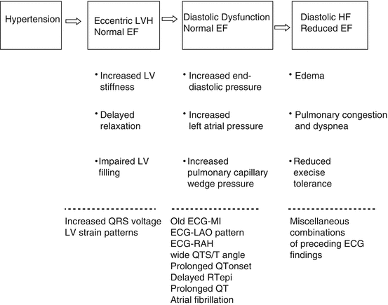
The block diagram in Fig. 7.1 is a schematic of the pathway of the evolution of LVH into left ventricular dysfunction and HF. The bottom section of the diagram lists ECG findings that can be expected in the course of the evolution. When early signs of diastolic dysfunction develop and the ejection fraction (EF) is still within normal limits, various ECG abnormalities that can be expected include transient AF, old ECG-MI, ECG signs of left atrial overload, wide QRS/T angle, delayed epicardial repolarization time (RTepi) and prolonged rate-adjusted QTend (QTea).


Fig. 7.1
A schematic showing ECG findings that can be expected (bottom section) in the evolution of hypertension to eccentric LVH, diastolic dysfunction and diastolic heart failure. LV left ventricular, EF ejection fraction, MI myocardial infarction, LAO left atrial overload, RAH right atrial hypertrophy, RTepi Epicardial repolarization time
Frequently used ECG-LVH criteria in prognostic evaluation are listed in Table 7.1 as common reference for this chapter. The following criteria were included: Sokolow-Lyon [5], Minnesota Code [6], Cornell voltage [7], Cornell product [8], Estes score [9] and Perugia score [10].
Table 7.1
Commonly used ECG criteria for left ventricular hypertrophy
Criteria [reference] | Variables | Criteria sets and limits |
|---|---|---|
Sokolow-Lyon (SL) [5] | RV5 + SV1 | 1. SL >3,500 μV (men and women) |
Minnesota Code MC 3.1 [6] | RI; RII; RIII; RaVL; RaVF; RV5 | 2A. Definite: R(I, II, III or AVF) >2,000 μV OR RaVL >1,200 μV OR RV5 > 2,600 μV |
2B. Borderline: 2A and (MC 4.1–4.3 or 5.1–5.3) | ||
Minnesota Code MC 3.1 + 3.3 | MC3.1 variables; RI; SL | 3A. Definite: MC 3.1 OR SL >3 500 μV OR |
RI >1,500 μV (men and women) | ||
3B. Borderline: 3A and (MC 4.1–4.3 or 5.1–5.3) | ||
Cornell voltage (CV) [7] | RaVL + SV3 | 4. CV >2,800 μV in men, >2,000 μV in women |
Cornell product (CP) [8] | CV•QRSdur. | 5. CP >240 μV.s (men and women) |
Estes score ≥4 (ES4) [9] (possible LVH) | RV5; RV6; RI; RII; RIII; RaVR; RaVL; RaVF; SI; SII; SIII; SaVR; SaVL; SaVF QRSdur; LV strain† | 6. RV5 or V6 ≥ 3,000 μV OR (R or S in any limb lead ≥3,000 μV) OR LV strain AND |
QRS >90 ms (men and women) | ||
Estes score ≥5 (ES5) (definite LVH) | Variables above and P’V1 | 7. RV5/6 ≥ 3,000 μV OR SV5/6 ≥ 3,000 μV OR R or S in any limb lead ≥2,000 μV OR LV strain |
Framingham score [9] | SL; RI; SIII; RaVL; SV1, V2; SL; LV strain | 8. Borderline: SL criteria OR RaVL >1,100 μV OR (RI + SIII) ≥2,500 μV or RV5/6 ≥ 2,500 μV OR SV1/V2 ≥ 2,500 μV |
9. Definite: borderline criteria + LV strain | ||
Perugia score [10] | SV3 + RaVL; | 10. Men: RaVL + SV3 > 2,400 μV O RLV strain pattern or Romhilt-Estes score ≥5; |
Left ventricular strain; | ||
Romhilt-Estes score | Women: RaVL + SV3 > 2,200 μV OR LV strain pattern OR Romhilt-Estes score ≥5; |
Several ECG models have been introduced for prediction of left ventricular mass (LVM) on a continuous scale (Echo-LVM) [11–13] including a model derived in the Cardiovascular Heart Study (CHS) [13]. The algorithms for LVM from the CHS model are listed in Table 7.2 for normal ventricular conduction, anterior/lateral MI and ventricular conduction defects according to the Minnesota Code. Ranking of the best individual variables in each model according to their partial R-square values revealed that body weight was a dominant predictor in normal conduction, inferior myocardial infarction (MI) and right bundle branch block (RBBB), QRS duration in left bundle branch block (LBBB) and anterior and lateral MI and JV6 amplitude in indetermined-type ventricular conduction defect (ICVD) (not shown). The correlation between ECG-LVM from this model and Echo-LVM was 0.54 in women and 0.51 in men. In CHS study ECG-LVM and Echo-LVM had about equally strong associations with overt and subclinical disease and with risk factors for left ventricular hypertrophy.
Table 7.2
ECG-LVH and risk of adverse outcome events from the Framingham observational studies and two intervention studies
Study | Population | Endpoint | ECG-LVH criteria | Adjusted risk estimates (95 % CI) |
|---|---|---|---|---|
Framingham [16] | 28–62 years; CVD-free at baseline | Fatal and non-fatal CVD events | Cornell Voltage (CV) as a continuous variable | Baseline Quartile 4 vs. Quartile 1: |
Women: 3.29 (1.78–6.09) | ||||
Men: 3.08 (1.87–5.07) | ||||
CV decrease vs. no change during follow up | ||||
Women: 0.56 (0.30–1.04) | ||||
Men: 0.46 (0.28–0.54) | ||||
CV increase vs. no change during follow up | ||||
Women: 1.61 (0.94–2.84) | ||||
Men: 1.86 (1.14–3.03) | ||||
Framingham [16]
Stay updated, free articles. Join our Telegram channel
Full access? Get Clinical Tree
 Get Clinical Tree app for offline access

|