The Third and Fourth Heart Sounds
PRETEST 
1. Describe a normal S3.
2. Describe an abnormal S3 and a right-sided S3.
3. What is a ventricular gallop, or gallop rhythm?
4. Describe a normal S4.
5. Describe an abnormal S4, a summation gallop, and a right-sided S4.
VENTRICULAR FILLING SOUNDS
The first and second heart sounds, S1 and S2, mark the beginning and end, respectively, of ventricular systole. In healthy individuals, these two sounds and their components are relatively easy to hear during auscultation and are best heard with the diaphragm of the stethoscope.
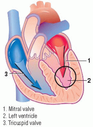
The two left ventricular diastolic filling sounds, S3 and S4, are sometimes heard over the mitral area. These sounds differ from S1 and S2 in that they’re low-frequency sounds produced by ventricular filling, rather than associated with valve closure.
THIRD HEART SOUND
Normal S3
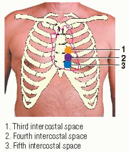
 Area producing S3 |
Occasionally, a physiologic third heart sound, S3, is heard during auscultation. This heart sound, considered normal in healthy individuals younger than age 20 and in athletic young adults, is caused by vibrations occurring during rapid, passive ventricular filling.
In patients older than age 40, an S3 is likely the result of heart failure or ventricular volume overload caused by valvular disease.
Early in diastole, after isovolumic relaxation, the mitral and tricuspid valves open and the ventricles fill and expand. In children and young adults, the left ventricle is normally compliant, permitting rapid filling. The left ventricle responds to this rapid filling with an abrupt change in wall motion that causes a sudden decrease in blood flow. These events generate vibrations that are responsible for the physiologic S3. The more vigorously the left ventricle expands, the greater the likelihood that an S3 will occur.
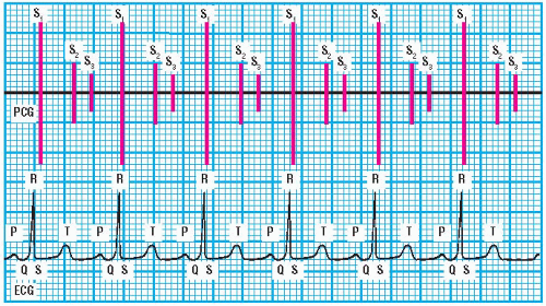 PCG and ECG showing physiologic S3 |
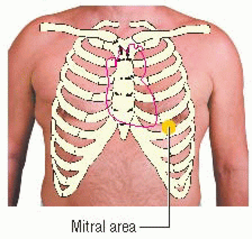 Auscultatory area for S3 |
A physiologic S3 is also commonly audible in patients with high-output conditions in which rapid ventricular expansion, caused by increased blood volume, is present. Anemia, fever, pregnancy, and thyrotoxicosis are conditions that cause rapid ventricular expansion, resulting in an S3. An S3 is also commonly heard in young, slender individuals during periods of excessive catecholamine release.
Auscultatory area and relationship to ECG
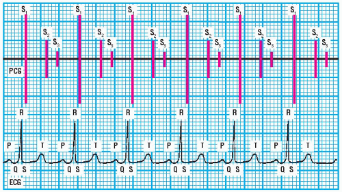
S3 is typically heard best with the bell of the stethoscope over the mitral area, and it can usually be palpated over the same area. It’s heard best during expiration when blood flow into the left ventricle is increased. (♦Sound 16) S3 is heard early in diastole and follows S2 by 0.14 to 0.20 seconds. The relationship of S3 to the electrocardiogram (ECG) waveform can be seen easily on a phonocardiogram (PCG). In the ECG, S3 occurs during the T-P interval just after the T wave. Normally, the S2-S3 interval is reliably constant.
Sound characteristics
S3 is usually heard best near the apex of the heart, over the mitral area. In some patients, it’s soft and faint in intensity and difficult to hear. In others, it may be loud and easy to hear. S3 has a short duration, and it may occur only intermittently during every third or fourth heartbeat.
S3 has a low pitch that’s heard best with the bell of the stethoscope. It usually has a dull, thudlike quality. Its timing is closely
related to S2, which is heard just after the T wave. S3 follows S2 by less than 0.20 seconds. (♦Sound 16)
related to S2, which is heard just after the T wave. S3 follows S2 by less than 0.20 seconds. (♦Sound 16)
 PCG and ECG showing abnormal S3 in a patient older than age 20 with a clinical condition |
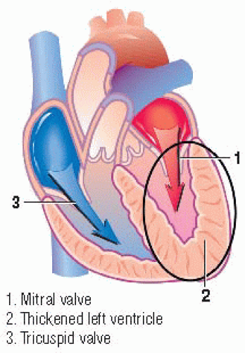 Area producing abnormal S3 |
Enhancing S3
Because S3 is associated with blood volume and velocity, it can be intensified by maneuvers that increase stroke volume such as elevating the patient’s legs from a recumbent position or having the patient exercise briefly or cough several times. A physiologic S3 usually disappears with maneuvers that decrease the venous return, such as having the patient sit up or stand.
 AUSCULTATION TIP
To enhance your ability to auscultate and palpate for an S3, place the patient in a partial left lateral recumbent position.
Abnormal S3
The S1, S2, S3 sequence is referred to as a ventricular gallop or gallop rhythm. (♦Sound 17) An abnormal S3 has the same sound characteristics, is heard over the same location, and has the same timing in relation to S2 as a physiologic S3. The differences between the two are related to the patient’s age, clinical condition, or both. Also, an S3 gallop rhythm usually persists despite maneuvers that decrease the venous return. (♦Sound 17)
An abnormal S3 is heard in conditions associated with increased blood volume and increased inflow velocity into the left ventricle. It is heard with or without an increase in ventricular diastolic pressure. Consequently, patients with mitral regurgitation and heart failure have an abnormal S3. This heart sound can also be heard during increased blood flow through the mitral valve.
 PCG and ECG showing pericardial knock |
 Auscultatory area for pericardial knock |
This occurs in patients with a ventricular septal defect, patent ductus arteriosus, or severe aortic regurgitation.


