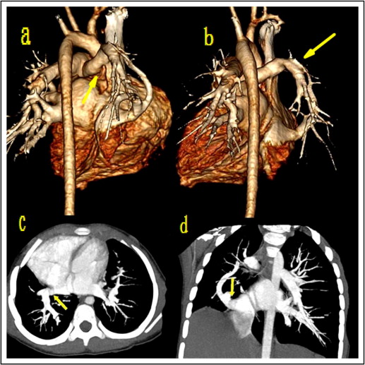
Scimitar syndrome is a relatively rare variety of congenital heart disease characterized by partial or complete anomalous pulmonary venous connection of the right lung into the inferior vena cava. There are virtually no reports of the use of 320-slice computed tomography in establishing the diagnosis. The investigators present a case of scimitar syndrome confirmed by 320-slice computed tomography.
Case Presentation
A 4-year-old female child was hospitalized for pneumonia. Physical examination disclosed normal sinus rhythm and normal results on auscultation. There was a small right hemithorax, decreased intensity of breath sounds on the right side, and occasional right-sided crepitations. Scimitar syndrome was demonstrated on 320-slice computed tomography ( Figure 1 ) , and the diagnosis was confirmed on cardiothoracic surgery. The patient’s postoperative course was uneventful.


