The Role of Intravascular Ultrasound in the Catheterization Laboratory


The Role of Intravascular Ultrasound in the Catheterization Laboratory


John McB. Hodgson

Suzanne Sorof


Intravascular ultrasound (IVUS) allows the direct visualization of coronary anatomy during diagnostic and interventional catheterization (1, 2, 3, 4, 5, 6). Unlike angiography, which depicts a silhouette of the coronary lumen, IVUS displays a tomographic, cross-sectional perspective. This facilitates direct measurements of lumen dimensions, including minimum and maximum diameter and cross-sectional area (7). By employing a timed pullback, length measures may be obtained (8). Ultrasound-derived measurements are more accurate than angiographic dimensions (9,10). In addition to luminal measurements, the ability of coronary ultrasound to image the soft tissues within the arterial wall enables the characterization of atheroma size, plaque distribution, and lesion composition (11, 12, 13). Accordingly, ultrasound can detect the presence or absence of structural abnormalities of the vessel wall after mechanical interventions, including dissections, tissue flaps, intramural hematomas, perforations, and irregular surface features (14, 15, 16, 17). Since intracoronary ultrasound was first performed in 1988, it has been instrumental to our understanding of coronary anatomy and pathophysiology and allowed detailed evaluation of interventional procedures. In this chapter, we explore the fundamental knowledge base related to IVUS in the catheterization laboratory.


INTRAVASCULAR ULTRASOUND DEVICES

Intracoronary ultrasound equipment requires two components: a catheter incorporating a miniaturized transducer, and a console containing the necessary electronics to reconstruct an ultrasound image. Catheters typically range in size from 2.9F to 3.5F, and a corresponding diameter of 0.96 to 1.17 mm. Two technical approaches to transducer design have emerged: mechanically rotated imaging devices and a multielement electronic array device. The mechanically rotated design requires an imaging sheath; the electronic design is inserted directly into the artery. Most systems use a monorail design to facilitate rapid catheter exchange.


ARTIFACTS AND LIMITATIONS

Mechanical transducers may exhibit variations in rotational speed, which arise from mechanical drag on the catheter driveshaft, creating nonuniform rotational distortion (NURD) and producing visible distortion. NURD is most evident when the driveshaft is bent into a small radius of curvature by a tortuous vessel; it is recognized as the circumferential stretching of a portion of the image
with a corresponding compression of the contralateral vessel wall. An additional artifact, transducer ring-down, appears in virtually all medical ultrasound devices. This artifact arises from acoustic oscillations in the piezoelectric transducer material and results in high-amplitude signals that obscure near-field imaging. In mechanical systems, this artifact may be merged with the imaging sheath artifact. In electronic array catheters, this artifact may be removed largely by mask subtraction. All intravascular imaging systems are vulnerable to the geometric distortion produced by oblique imaging. Thus, when the ultrasound beam interrogates a plane not orthogonal to the vessel walls, an artery with a circular lumen appears elliptical in shape. Some transducer designs position the guide wire external to the transducer, thereby introducing an obligatory wire artifact. In general, higher frequency transducers have a lower penetration depth; in practice, this is not usually an issue for coronary imaging, but may become evident if peripheral arterial imaging is attempted. Alternative catheters with reduced frequency are used for large-vessel peripheral imaging and for intracardiac examination.


SAFETY OF CORONARY ULTRASOUND

Although IVUS requires intracoronary instrumentation, initial studies conducted during diagnostic catheterization demonstrated few serious untoward effects (18, 19, 20). The imaging transducer transiently can occlude the coronary when advanced into a tight stenosis or a small distal vessel, but patients generally do not experience chest pain if the catheter is promptly withdrawn. Preinstrumentation nitroglycerine is advised to prevent spasm. In interventional practice, operators safely have used coronary ultrasound after most types of procedures, including balloon angioplasty and stent deployment. Despite the relative safety of coronary ultrasound, any intracoronary instrumentation carries the potential risk of intimal injury or acute vessel dissection. Although many centers use IVUS during diagnostic catheterization, most laboratories limit credentialing for intravascular imaging procedures to personnel with interventional training. In the unlikely event of intimal disruption, this safety measure ensures that the necessary personnel and equipment are immediately available to initiate appropriate interventional corrective action.


QUANTITATIVE LUMINAL MEASUREMENTS

Diagnostic and interventional practitioners routinely use luminal measurements to evaluate the severity of stenoses, determine the size of the “normal” reference segment, and assess the gain in lumen size achieved by revascularization. Comparisons of vessel dimensions by angiography and IVUS generally reveal a limited correlation, particularly for vessels with an eccentric luminal shape (21), presumably owing to the inability of angiography to accurately portray the complex, irregular cross-sectional profiles of atherosclerotic vessels. Poor correlations have been found between angiographic and IVUS evaluation after balloon angioplasty due to the complex dissections that occur (22). In general, angiography overestimates lumen dimensions compared to IVUS, even after symmetric stent implantation (23). By performing a timed or calibrated pullback through the vessel, a third dimension of information can be collected (length), thus allowing the calculation of lumen, vessel, and plaque volume (24). This information has been instrumental in evaluating the mechanism of different interventional techniques, the pathology of restenosis, and the effect of drug therapy aimed at treating atherosclerosis.


ANGIOGRAPHICALLY UNRECOGNIZED DISEASE

IVUS commonly detects atherosclerotic abnormalities at angiographically normal coronary sites (25, 26, 27, 28). The long-term implications of these findings remain uncertain. However, Little et al. (28) have demonstrated that plaques with minimal to moderate angiographic narrowing are the most likely to rupture and cause acute coronary syndromes. Accordingly, the presence of angiographically occult coronary disease may have important prognostic significance. Studies are currently under way to determine the predictive value of IVUS in determining the prognosis of patients with coronary disease. Several studies have shown that plaque burden or lumen size in the left main coronary may be an indicator of the behavior of the coronary vasculature, and that the left main disease seen on IVUS predicts adverse cardiac events (29,30).

Using volumetric intracoronary ultrasound, minor changes in plaque and lumen volume can be detected reliably. By comparing baseline and repeat studies, the effects of drug therapy on atheroma can be assessed (31, 32, 33). Due to the precise measures, such methodology allows pharmaceutical studies to be completed using far fewer patients than necessary if only clinical endpoints are collected (34,35).


LESIONS OF UNCERTAIN SEVERITY

Despite thorough radiographic examination using multiple projections, angiographers commonly encounter lesions that elude accurate characterization. Coronary atherosclerosis can be associated with vessel remodeling and dilatation, thus the angiographic appearance of the vessel may be normal despite a significant accumulation of plaque. Lesions of uncertain severity often include ostial lesions and moderate stenoses (angiographic severity ranging from 40% to
70%) in patients whose symptomatic status is difficult to evaluate. For these ambiguous lesions, ultrasound provides tomographic measurements, enabling the quantification of the stenosis independent of the radiographic projection (36,21). Bifurcation lesions are particularly difficult to assess by angiography because overlapping side branches often obscure the lesion (37). Intravascular lesion cross-sectional areas have shown a statistically significant, although weak, correlation with noninvasive stress imaging studies and other measures of stenosis severity (38, 39, 40, 41, 42). In general, lumen areas less than 3 to 4 mm2 correlate with a positive stress study. Our own longitudinal study of 36 patients in whom IVUS was performed to assess lesion significance and for whom treatment was deferred showed a 14.3% major adverse cardiac event rate at a mean follow-up of 18.8 months (Fig. 5A.1). Generally, however, functional measures such as fractional flow reserve may be better suited for assessing the prognostic significance of intermittent lesions.

Because the severity of left main disease is of critical importance for properly determining patient treatment, intravascular imaging of this vessel deserves special mention. Several studies have documented the negative impact of left main disease discovered by IVUS on patient prognosis (29,30,43, 44, 45, 46). In one study of 122 consecutive patients who had left main imaging and were treated medically, lesions in the left main having lumen diameters less than 3 mm clearly predicted a higher incidence of subsequent cardiac events (30).


CARDIAC ALLOGRAFT DISEASE

The identification of atherosclerotic lesions in cardiac allograft recipients represents a particularly challenging task (26,47). These patients may have diffuse vessel involvement that, for reasons already enumerated, conceals the atherosclerosis from angiography. Many large transplant centers now routinely perform IVUS as an annual catheterization in all cardiac transplant recipients. Studies have revealed two pathways to transplant-associated atherosclerosis, with some patients receiving atherosclerotic plaques from the donor heart, whereas others develop immune-mediated vasculopathy (47,48).


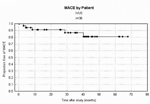
Figure 5A.1. Kaplan-Meyer curve displaying the rate of major adverse cardiac events (MACE) in patients treated medically after IVUS evaluation of intermediate lesions. Sixteen of these lesions were in the left main coronary.


INTRAVASCULAR ULTRASOUND AND RESTENOSIS

The relatively poor correlation between angiographic and ultrasonic dimensions after percutaneous coronary intervention (PCI) raises the issue of whether poor long-term results represent recurrence of disease or an inadequate initial procedure (49). Several multicenter clinical trials have shown that certain findings on ultrasound, such as minimal lumen area and plaque burden, can predict restenosis after intervention. The Guidance by Ultrasound Imaging for Decision Endpoints (GUIDE) trial evaluated the predictive value of intravascular imaging after balloon angioplasty. A residual plaque burden in the lesion of >65% or a minimal lumen area of <2 mm2 was associated with a higher rate of restenosis (50). A careful longitudinal study of balloon angioplasty and directional atherectomy documented that the primary mechanism of restenosis was vessel contraction (negative remodeling) (Serial Ultrasound Analysis of Restenosis [SURE] trial) (51). Stent placement abolishes this negative remodeling; however, it stimulates intimal hyperplasia inside the stent (52). The major predictor of in-stent restenosis has been shown repeatedly to be the final
minimal stent area. Areas over 8 to 10 mm2 are generally associated with target lesion revascularization rates of 10% or less (53, 54, 55, 56). Although inadequate minimal stent area is the predominant factor associated with in-stent restenosis lesions, nearly 5% of cases are found to have significant mechanical implantation abnormalities (57). Many experts recommend that all cases of in-stent restenosis be interrogated by IVUS to define the mechanism and guide therapy.

Intracoronary brachytherapy is an effective treatment for in-stent restenosis, resulting in a significant retardation of neointimal hyperplasia formation. Although intravascular imaging was helpful in developing the brachytherapy delivery systems and dosing strategies, it is not generally felt to be necessary for the guidance of commercial brachytherapy delivery systems (58,59).


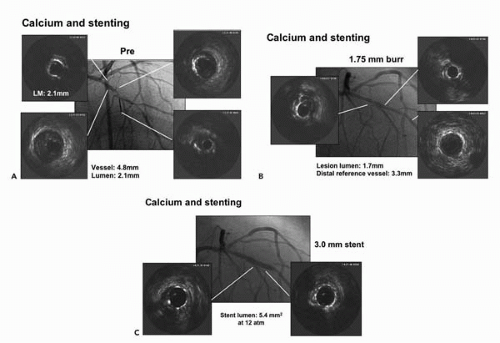
Figure 5A.2. Case study of a severely calcified left anterior descending (LAD) lesion. The IVUS catheter would not pass the lesion initially (A). The left main (LM) is diffusely calcified, with a lumen diameter of only 2.1 mm (upper left panel). In the mid LAD, the vessel (EEM) diameter is 4.8 mm (lower left panel). In the lesion, extensive calcium is appreciated (lower right). Following treatment with a 1.75 mm rotational atherectomy burr (B) the IVUS catheter is able to be passed into the distal vessel (lower right panel). The distal reference vessel diameter is 3.3 mm, allowing selection of a 3.0 mm stent. Following 3.0 mm stent implantation, the angiographic appearance is excellent (C). IVUS, however, shows that the stent lumen area is still relatively underexpanded (5.4 mm2 compared with a nominal area of 7.1 mm2 for a 3 mm stent). Angiographically guided stent size selection would have been even smaller.


BALLOON ANGIOPLASTY

Early intravascular studies determined the mechanism of balloon angioplasty (15,60, 61, 62). The primary mechanism of lumen enlargement is overstretching of the adventitia, with axial plaque redistribution away from the lesion center. Specific predictors of restenosis following balloon percutaneous transluminal coronary angioplasty (PTCA) include residual plaque area and minimal lumen area (50,63, 64, 65). After intravascular imaging confirmed marked occult atheroma in the “reference” segments surrounding target lesions, several investigators explored the use of oversize balloons to enhance balloon angioplasty (66, 67, 68, 69, 70, 71, 72). This “provisional-stent” strategy resulted in excellent angiographic and clinical outcomes (73). Approximately 50% of lesions could be treated effectively in this way, with the other 50% requiring stenting to achieve optimal results. Although once proposed as means of avoiding costly stents, this strategy has not been widely adapted.


TABLE 5A.1. IVUS- VERSUS ANGIOGRAPHICALLY-GUIDED PCI TRIALS


Year


Study


Study Design


Primary Results


Points of Interest


1995


Colombo (92)


Single-center registry 359 patients; IVUS after angiographically adequate stent implantation


30% of stents placed had optimal expansion after angiographic guidance (CSA >60% of reference). After IVUS guidance, this increased to 96%; subsequent 6-month SAT was 1.6% on antiplatelet therapy only.


IVUS guidance resulted in a 26% increase in minimal stent area (MSA). Study led to now-routine process of high-pressure stent implant without postprocedure coumadin.


1997


Albiero (115)


Case control (two sites) of 346 patients. IVUS- versus angiographically guided stent


Early after stent implant, restenosis is less with IVUS guidance (9.2% IVUS vs. 28.3% angio; p = 0.04). No difference at late follow-up (22.7% vs. 23.7%).


Acute results of stent implantation can be optimized with IVUS.


1997


Stone (66) CLOUT


Multicenter registry of 102 patients. IVUS-guided PTCA after angiographically adequate result


IVUS-measured EEM diameter in reference vessel used to define oversized balloon diameter. Increased balloon size in 73% of lesions. No increase in dissections after use of IVUS-determined oversize balloon.


Due to positive remodeling, oversized balloons (B:A ratio 1:3) may be used safely for PTCA. IVUS is needed to assess degree of remodeling (not apparent angiographically).


1998


de Jaegere (116) (MUSIC)


Multicenter registry of 161 patients. IVUS guidance of stent implantation


Stents implanted using IVUS-guided criteria associated with a 6-month TVR rate of 4.5% and angiographic restenosis rate of 9.7%. Antiplatelet agents only with optimal IVUS result. No acute Q-wave MI, one CABG, 1.3% SAT.


Tested hypothesis that criteria-driven IVUS guidance could improve restenosis after stenting. Compared to contemporary stent studies, this had lowest reported TVR rate.


1998


Schiele (119) (RESIST)


Randomized multicenter trial of 155 patients. IVUS-versus angiographically guided stent


80% achieved IVUS criteria. Stent CSA in IVUS group 20% larger. Restenosis at 6 months (22.5% IVUS versus 28.8% angio; p = 0.25). Lumen area by IVUS only predictor of restenosis.


IVUS group had larger 6-month MSA. Study was underpowered for restenosis endpoint.


1999


Schroeder (69)


Single-center registry of 252 consecutive patients. IVUS-guided PTCA


PTCA with balloon sized 1:1 with lesion EEM resulted in a low MACE rate (14%) and low angiographic restenosis rate (19%). Only 2% received stents.


Aggressive PTCA strategy based on vessel remodeling. Many dissections were left untreated and were not associated with adverse events.


1999


Abizaid (70)


Single-center registry of 284 consecutive patients; IVUS-guided provisional stenting


PTCA with balloon sized 1:1 with lesion EEM. Stents placed for inadequate results. 47% of procedures successful with balloon only. Overall 1-year TLR 12% (IVUS-guided PTCA lesions: 8%, stented lesions: 16%.)


Aggressive PTCA strategy with more liberal use of stents for dissections than in the Schroeder study. Clinical results after IVUS-guided balloon-only are excellent.


2000


Frey (67) SIPS


Randomized single-center trial of 291 consecutive patients. IVUS-guided versus angiographically guided PCI


Stent use encouraged only for inadequate PTCA result and was similar in two groups (47.7% vs. 49.0%).
6-month restenosis: 29% IVUS versus 35% angio; p = 0.42. 2-year TLR: 17% versus 29%; p = 0.02.


Clinical outcome at 2 years improved by routine IVUS guidance of PCI. IVUS strategy was cost effective.


2000


Fitzgerald (117) CRUISE


Case control (multicenter) study of 525 patients. IVUS-versus angiographically guided stent


Acute MSA: 7.78 mm2 IVUS versus 7.06 mm2 angio; p <0.001.
9-month TLR: 8.5% IVUS versus 15.5 angio; p <0.05.


Clinical outcomes at 9 months improved by routine IVUS guidance of Palmaz-Schatz stent implantation.


2000


Russo (118) AVID


Randomized multicenter trial of 759 patients. IVUS-versus angiographically guided stent


Acute MSA: 7.54 mm2 IVUS versus 6.94 mm2 angio; p <0.01.
12-month TLR significantly better in IVUS-guided graft or smaller vessel (<3.25) lesions.


Final report not yet publicized. TLR reported as 4.9% IVUS versus 10.8% angio in protocol-compliant patients.


2001


Mudra (120) OPTICUS


Randomized multicenter trial of 550 patients. IVUS-versus angiographically guided stent


6-month restenosis: 24.5% IVUS versus 28.8% angio; p = 0.68.
12-month MACE: Relative risk 1.07; p = 0.71. 12-month TLR: Relative risk 1.04; p = 0.87.


Only neutral randomized trial. Acute gain greater in IVUS group (2.07 mm2 versus 1.93 mm2; p <0.001).


2003


Oemrawsingh (123) TULIP


Randomized single-center trial of 144 patients. IVUS-versus angiographically guided stents


Stenosis >20 mm long; 6-month restenosis: 23% IVUS versus 45% angio; p = 0.008.
12-month TLR: 10% versus 23%, p = 0.018.


Clinical outcome improved with IVUS guidance of long-lesion stenting.


2003


Schiele (72) BEST


Randomized multicenter trial of 254 patients. IVUS-guided PTCA versus angiographically guided stent


Noninferiority design 44% of IVUS PTCA group received stents. 6-month restenosis: 16.8% IVUS versus 18.1% stent, noninferior.


IVUS-guided PTCA with provisional stenting not inferior to routine angiographically guided stent implantation.

Only gold members can continue reading. Log In or Register to continue

Stay updated, free articles. Join our Telegram channel

Sep 23, 2016 | Posted by in CARDIOLOGY | Comments Off on The Role of Intravascular Ultrasound in the Catheterization Laboratory

Full access? Get Clinical Tree

Get Clinical Tree app for offline access