THE PULMONARY CIRCULATION: BRINGING BLOOD AND GAS TOGETHER

7


THE PULMONARY CIRCULATION


BRINGING BLOOD AND GAS TOGETHER


The functions of the pulmonary circulation


So far, we have considered how the respiratory system transports gas between the atmosphere and the alveoli. In this chapter we will consider how blood and gas are brought together so that gas exchange can take place between them. This is a complex process: in order for all blood to undergo gas exchange, virtually the entire cardiac output is directed through the lungs, and brought close to gas within the alveoli. Furthermore, it is very important that ventilation (gas flow) and perfusion (blood flow) to a given area of the lung are matched. There is no point in directing air to areas of the lung that have little blood flow, and conversely there is no point in perfusing an area of lung with little or no ventilation. This matching of ventilation and perfusion is vital to maintaining adequate oxygenation and carbon dioxide removal from the blood.


In this chapter we will consider the anatomy of the pulmonary circulation, and compare it to the systemic circulation. We will then consider the factors that influence the flow of blood to different regions of the lungs. Finally, we will consider the very important issue of ventilation/perfusion matching: the way in which air and blood are brought together in order to achieve optimal gas exchange of oxygen and carbon dioxide.


The anatomy of the pulmonary circulation


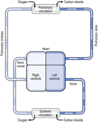
In humans the circulation behaves in many ways as if it consisted of two parts. In the systemic circulation the left ventricle pumps oxygenated blood through the organs and tissues of the body, where oxygen is removed and carbon dioxide added before the blood returns to the right atrium. In the pulmonary circulation the right ventricle pumps deoxygenated blood through the lungs, where oxygen is added and carbon dioxide is removed (Fig. 7.1). Of course, the two parts of the circulation work in series to form a single circuit of blood, but there are a number of differences between the pulmonary and systemic circulations which reflect their different functions.


First, virtually the entire cardiac output is directed through the pulmonary circulation. This means that, at any one time, there is as much blood flowing through the lungs as through all the other organs and tissues in the body put together.


Second, the purpose of the pulmonary circulation is to bring blood and air into very close contact in order to allow gas exchange to take place. This requires a very thin separating membrane and for this reason the pressure in the pulmonary circulation needs to be very low compared to the systemic circulation. If the pressure in the pulmonary circulation were higher, it could cause fluid to leak from the pulmonary capillaries into alveoli. In fact, the pressure in the pulmonary artery is about 25/10 mmHg, compared to 120/70 mmHg in the systemic circulation. The pulmonary circulation is therefore a low-pressure, high-flow system, which implies it has a low resistance. The components of the pulmonary circulation differ from their systemic counterparts in a way that reflects this.


The right ventricle


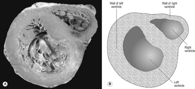
Every minute, the same volume of blood – about 5 L at rest – flows through both the right ventricle and the left ventricle. However, the two ventricles look very different (Fig. 7.2). The left ventricle has a thick, muscular wall that takes up most of the cross-section of the heart. The right ventricle has a much thinner wall, about one-third of the thickness of the left, and to accommodate the muscle of the left ventricle the right almost seems to be ‘wrapped around’ the left. Why is there such a difference between the two?


The reason is that the left ventricle pumps blood into the systemic circulation, which has a high resistance and which operates at a relatively high pressure. If cardiac output increases, for example during exercise, this pressure can increase even more, which means that the left ventricle needs a thick, muscular wall to produce these high pressures. On the other hand, the right ventricle pumps blood into the pulmonary circulation, which has a very low resistance and which operates at a low pressure, always less than that in the systemic circulation. Furthermore, as we shall see later, if cardiac output increases the pulmonary artery pressure does not increase very much. For this reason, the right ventricle needs only a relatively thin muscular wall in comparison to the left.


Pulmonary blood vessels


Pulmonary blood vessels are very different from their systemic counterparts. The larger vessels have much thinner walls, reflecting the lower blood pressure they need to withstand: for example, the thickness of the wall of the pulmonary artery is only about one-third of the thickness of the aorta.


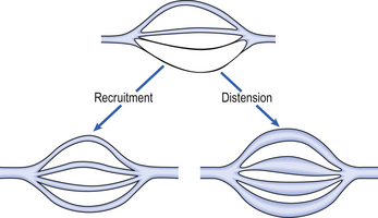
As well as having thinner walls, the pulmonary vessels are much more distensible than systemic arteries, which is important in keeping pulmonary blood pressure low during systole and in the face of increases in cardiac output. In circumstances such as exercise, cardiac output can increase from its normal 5 L per minute to as much as 25 L per minute. In order to keep pulmonary blood pressure low, the pulmonary circulation is able to reduce its resistance to even lower values than normal. It does so by two mechanisms, illustrated in Figure 7.3.


The anatomical position of the capillaries and their function in gas exchange means that they are different from their systemic counterparts. The density of the capillary network in the alveolar walls is extremely high so that efficient gas exchange can take place. In fact, there are so few cells between the capillaries in the alveolar walls that the alveolar circulation behaves almost like a film of blood flowing around the alveoli. At rest, blood flows through an alveolar capillary in about 0.8 seconds, which is about three times longer than the time needed for oxygenation of mixed venous blood.


There is very little space between the blood in the pulmonary capillaries and the air in the alveoli. In fact, for the most part, the only cells separating blood from air are the endothelial cells of the pulmonary capillary and the epithelial type I cells of the alveolar wall (Fig. 2.9). This is clearly very important in allowing the most efficient transfer of gases between the alveoli and the blood (see Chapter 6).


However, because the pulmonary capillaries are very thin-walled and lie in the alveolar walls they can be readily influenced by changes in the gas pressure within the alveoli. Increased alveolar gas pressure can compress the capillaries, with a consequent increase in capillary resistance and reduction in blood flow, which can affect the distribution of blood flow within the lungs (see below). This is very different from the conditions affecting systemic capillaries, which are supported by surrounding tissues.


In contrast to the situation in the systemic circulation, the pressure in the pulmonary veins exerts a considerable influence over the pressure in the pulmonary arteries. This is another consequence of the low pressures in the pulmonary circulation.


In the systemic circulation, the pressure at the precapillary sphincters, which regulate the blood flow through the capillary beds, is about 90 mmHg, very much higher than the pressure at the venous end of the capillary beds. The difference in pressure between the arterial and venous ends of a capillary bed is called the driving pressure and flow is dependent on this. Because the venous pressure in the systemic circulation is so much less than the arterial pressure, changes in venous pressure do not make large changes to the driving pressure.


However, in the pulmonary circulation the difference between pulmonary arterial and venous pressures is much less, and a relatively small change in venous pressure can make a considerable change to the driving pressure. To keep a constant driving pressure the pulmonary artery pressure has to increase. In certain pathological situations this can eventually lead to failure of the right ventricle (cor pulmonale; see below).


The bronchial circulation


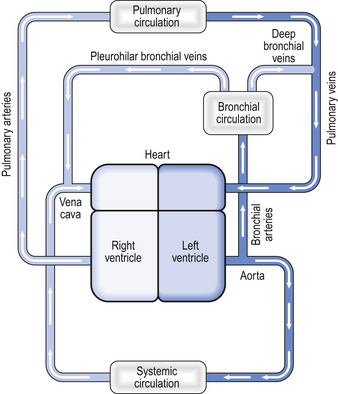
Not all the blood flowing into the lungs does so via the pulmonary artery – a small volume of arterial blood flows through the bronchial circulation (Fig. 7.4). The bronchial circulation supplies blood to the airways and to the lung parenchyma, although it is not essential to their survival; during a lung transplant the bronchial circulation is not reconnected, and this apparently does not produce any serious ill effects. Blood flowing through the bronchial circulation does not pass through the alveolar capillaries and therefore does not take part in gas exchange.


The bronchial arteries arise from the aorta. Bronchial vessels supply blood to the lower trachea, the bronchi, and to the smaller airways as far as the respiratory bronchioles. Blood from the proximal part of the bronchial circulation around the bronchi drains via the pleurohilar bronchial veins into the azygous vein and into the superior vena cava. This blood is effectively part of the systemic circulation in that it flows from the aorta to the vena cava.


However, the blood from the more distal parts of the bronchial circulation drains via the deep bronchial veins into the pulmonary circulation. This blood therefore forms a shunt (see below). In other words, the blood arises from the aorta, but instead of draining into the right-hand side of the circulation, the partly deoxygenated blood of the deep bronchial veins drains into the oxygenated blood that has passed through the alveolar capillaries. The resulting mixture of blood therefore has an oxygen content less than that of the original pulmonary artery blood. The significance of this will be discussed later in the chapter.


Matching ventilation and perfusion


In an ideal pair of lungs all the alveoli would be supplied with equal volumes of air of uniform gas composition during inspiration. Also, all the alveoli would be ideally supplied with the same flow of mixed venous blood. The ventilation and perfusion of all parts of these ideal lungs would therefore be optimally matched, and optimal gas exchange between the blood and the alveoli would take place.


However, in real lungs this is not the case. Per unit of lung volume, ventilation and perfusion both tend to be greater at the bases of the lungs compared to the apices. Nevertheless, for most of the lung tissue the two tend to be fairly optimally matched. This means that the ratio of ventilation to blood flow, the ventilation/perfusion ratio, or image ratio, varies by a relatively small amount throughout the lungs.


In order to see how ventilation/perfusion matching takes place we will first look at the way in which the blood flow is distributed throughout the lungs, then we will see how this is matched with ventilation before looking at how regional variations in the image ratio in the lungs affects arterial blood gases.


Distribution of blood flow through the lungs


As we have just seen, the distribution of blood to different regions of the lungs is not uniform but varies considerably. Furthermore, the blood flow to different parts of the lungs tends to be directed towards maintaining as normal a image ratio as possible.


In the systemic circulation, blood flow through organs is almost entirely determined by high-resistance arterioles that regulate the blood flow through capillary beds. The arterioles in the pulmonary circulation do not have a high resistance and play only a small role in determining the blood flow to different parts of the lungs. The distribution of blood flow through the lungs is influenced instead by a number of different factors, including gravity, alveolar gas pressure, hypoxic pulmonary vasoconstriction and, to a lesser extent, the nervous control of blood vessel resistance.


Gravity


The diastolic blood pressure in the systemic circulation is about 80 mmHg, which is enough pressure to raise a column of water by a height of over a metre. In other words, there is more than enough pressure to carry blood from the heart up to the head. However, in the pulmonary circulation the diastolic blood pressure is about 12 mmHg, enough pressure to raise a column of water about 15 cm. In other words, there is only just enough pressure to pump blood from the right ventricle up to the lung apices. On the other hand, at the lung bases the blood pressure in the pulmonary circulation is equal to the pressure generated by the right ventricle plus the hydrostatic pressure of a column of blood extending up to the heart. Because the pressure generated by the right ventricle is not very high, this extra hydrostatic pressure makes a very significant difference. Thus there is a very considerable difference in arterial blood pressure between the bases and the apices of the lungs owing to gravity. In other words, gravity tends to direct blood towards the lung bases.


Case 7.1   The pulmonary circulation: bringing blood and gas together: 2


What causes a pulmonary embolus and how can it be diagnosed?


A pulmonary embolus occurs when something, usually a thrombus (blood clot), occludes part of the pulmonary artery tree. Generally the thrombus forms in the veins of the pelvis or lower limb, and part of that thrombus or the whole thrombus may dislodge and pass through the vena cava, through the right atrium and ventricle and into the pulmonary artery. The thrombus finally lodges in a branch of the pulmonary artery, occluding it. The segment of lung tissue supplied by the obstructed artery has a reduced blood supply (although it often receives some blood – remember the bronchial circulation) and may finally infarct.


In a small number of cases (probably less than 10% of the total number of pulmonary emboli) the thrombus does not form in the veins of the pelvis or leg but forms in the heart. This may be as a result of atrial fibrillation, in which the atria of the heart do not beat properly, or thrombus may form on a part of the myocardium which has infarcted. Very occasionally the embolus is not formed from thrombus but from other substances, such as fat or amniotic fluid.


Conditions that lead to the formation of thrombus in the pelvic and lower limb veins include prolonged immobility, lower limb or pelvic fractures, abdominal surgery, pregnancy, the presence of cancer and clotting abnormalities. Mrs Dodds had two of these risk factors, including immobility (she was bedridden) and a lower limb fracture. If the thrombus forms in the lower limb it may become swollen and painful, which is why the doctor examined Mrs Dodds’ legs. If thrombus occurs in the lower limbs it usually occurs in the deep veins in the muscle, rather than the veins near the skin. Hence the condition is usually called deep venous thrombosis (DVT). Several cases have been reported of patients suffering DVTs and pulmonary emboli following the prolonged immobility that occurs during a long-distance flight, sometimes in rather cramped conditions. The true incidence of this so-called ‘economy class syndrome’ is yet to be established, however.


Small emboli in the lungs cause no symptoms and no haemodynamic problems and go unnoticed. Larger emboli, particularly if they result in pulmonary infarction, can cause clinical symptoms, including pleuritic chest pain and sometimes haemoptysis. Very large emboli are a medical emergency (see Box 4).

Only gold members can continue reading. Log In or Register to continue

Stay updated, free articles. Join our Telegram channel

Jun 18, 2016 | Posted by in RESPIRATORY | Comments Off on THE PULMONARY CIRCULATION: BRINGING BLOOD AND GAS TOGETHER

Full access? Get Clinical Tree

Get Clinical Tree app for offline access