Chapter 13 The history of respiratory physiology
 That breathing was essential for life was clear to the ancient Egyptian civilisations 5000 years ago, but the reasons for this were unknown.


This chapter is of necessity only a brief overview of the subject, and ends around 100 years ago, when the explosion of scientific progress makes the subject too large for such a short account. Significant advances in respiratory physiology in the last 100 years are reported in the other chapters of this book, and the reader interested in the history of this period is referred to more authoritative accounts.1,2,3 For more general information on the history of respiratory physiology numerous recent sources (by historical standards) are available.4,5,6
Ancient Civilisations
Egyptian Physiology7
Medical papyri. Many Egyptian papyri are concerned with medical topics, mostly descriptions of pragmatic ‘recipes’ for the treatment of a multitude of specific conditions.7
The longest and best known of the medical papyri is the Ebers Papyrus,8,9 which dates from about 1534bc, and is accepted as being a compilation of various earlier works. The Ebers Papyrus is unique in containing a section on physiology, including comments on respiration. The overall purpose of respiration is described thus:
As to the air that penetrates into the nose. It enters into the heart and the lung. They are those which give air to the entire body.9
Further sections include detailed descriptions of specific numbers of metu conducting ‘moisture and air’ to many parts of the body. These metu seem to mostly relate to blood vessels but also probably included such structures as tendons, muscles and the ureters. At first, this primitive view of anatomy is surprising considering the embalming abilities of ancient Egyptians, though in practice, embalming was carried out using very small inconspicuous incisions that would have revealed very little internal anatomy. Two metu are described to each ear, through which ‘the breath of life enters into the right ear and the breath of death enters into the left’,9 illustrating the ‘magical’ aspect of medicine at the time.
Ancient Greece
Greek writers were primarily philosophers, but they were also outstanding physicians, with one of their number, Hippocrates, forming a school that is now widely attributed with the foundation of modern medical conduct. Early Greek philosophers such as Anaximenes (570bc–?) clearly stated that ‘pneuma’ or air was essential to life,6 but in contrast to this correct observation, Alcmaeon reportedly claimed that goats breathed through their ears and that some air passed from the nose directly to the brain.4 Empedocles (495–435bc) disputed many of Alcmaeon’s writings, suggesting instead that breathing occurred through the skin, and that blood flow from the heart was tidal in nature, ebbing and flowing to and from the heart. Empedocles successfully combined physiology and philosophy in his description of the ‘innate heat’ in the heart, which was closely related to the soul, and which was distributed throughout the body by the heart. This concept of heat generation within the heart gained acceptance throughout the ancient Greek period, and was to remain at the centre of respiratory physiological ideas for about 1000 years.
The writings of Plato, Aristotle and the Hippocratic school only rarely directed their attention to respiration, but their contribution to scientific method and thinking was enormous. Subsequent philosopher-physicians adopted a more scientific approach to investigating physiology. At this time, dissection became widely practised, sometimes in public, and on both animals and humans. Animal vivisection also undoubtedly took place, and there are even disputed reports of human vivisection of criminals.4 Herophilus (circa 325bc) distinguished between arteries and veins, and, along with Aristotle, asserted that they contained air. Erasistratus (304–250bc), more widely renowned as the father of philosophy, was the first to apply scientific principles to explain breathing. His view was that air was taken into the lungs and passed to the heart along the pulmonary artery. In the heart, air was converted into a ‘vital spirit’ that was distributed to all parts of the body by the arteries, whilst the brain further converted the vital spirit into ‘animal spirit’ which travelled down the hollow nerves to activate muscles. Erasistratus seemed to understand that heart valves only allowed flow to occur in one direction, but failed to apply this knowledge to elucidate the transport of vital spirit or blood around the body. After Erasistratus, Greek interest moved away from medicine to philosophy and the physical sciences, and the progression of physiological knowledge halted for about 400 years.
Roman Medicine and Galen (129–199AD)
By the age of 28 years Claudius Galen was physician to the gladiators of Pergamun, and 12 years later became physician to the Roman emperor Marcus Aurelius. He wrote many works on anatomy and physiology, many of which still exist in modern form including two with much material on respiration On the usefulness of the parts of the body and On the use of breathing.10,11 Galen’s work provides the first direct evidence of experimentation and the application of clinical observations to explain physiology.
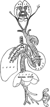
Galen’s system of physiology and anatomy. In Galen’s descriptions, food was processed in the gut before being used by the liver to produce blood, which passed to the right heart. Much of this blood flowed into the pulmonary artery to nourish the lung, whilst the remainder passed across invisible pores in the inter-ventricular septum, to be combined with ‘pneuma’ brought from the lung via the pulmonary vein (Figure 13.1). In the left heart, the pneuma instilled the blood with vital spirit that was circulated to the body and brain as described by Erasistratus.
Experiments on respiration. Galen’s experiments provided mixed results:
Galen’s conclusion from this study is contradictory: ‘Hence it is clear that the arteries all through the animal draw in the outer air very little or not at all’. Modern views of this experiment are that the ox-bladder was unlikely to be air tight, or that Galen’s assistants must have removed the bladder to allow the boy to breathe easily when their master was not directly supervising the experiment.11
There was also a darker and more controversial side to Galen. He is widely believed to have been conceited, dogmatic and abusive of those criticising him.6 On the usefulness of the parts of the body contains several prolonged and personal refutations of the ideas of his predecessors, for example, accusing ‘Asclepiades, wisest of men’ of making errors ‘no child would fail to recognise, not to mention a man so full of his own importance’.
After Galen
Preservation of knowledge now fell to scholars of the Byzantine and Arabic empires. The latter embraced Galen’s ideas with enthusiasm and translated many Greek works into Arabic, almost certainly adding their own refinements as they did so. The greatest of these Arabic scholars was Avicenna (circa 980–1037) whose canon was an impressive document pulling together and classifying all the available medical knowledge of the time, creating what has been described as a popular medical encyclopaedia of the medieval period. Some years later, Ibn Al Nafis13 (1210–1288), a prolific Arabic writer on many subjects, studied Avicenna’s writings and wrote his own treatise Sharh Tashirh Al-Qanun (Commentary on the Anatomy of the Canon of Avicenna). In this he challenged Galen’s scheme of pores in the inter-ventricular septum through which blood passed, and instead suggested that blood passed through the lung substance where it permeated with the air.5,13 This was an early breakthrough in explaining the true nature of the pulmonary circulation, but Ibn Al Nafis’ work did not become well known for many more centuries.
The Renaissance
In the 12th and 13th centuries, scholastic pursuits began again with the foundation of many European universities, firstly Oxford, Cambridge and Bologna closely followed by Paris, Naples and Padua. Soon, many of the ancient documents were translated from Greek or Arabic into Latin, and human dissection began to be performed after many centuries of interdiction by the Pope. Knowledge of anatomy again began to advance, though interest in the function of the body only began again with Leonardo da Vinci in the 15th century.
Leonardo Da Vinci (1452–1519)4
Leonardo exemplified the Renaissance trend for combining art with science. His anatomical drawings are both extensive and ingenious, being mostly surrounded by extensive explanatory notes.14,15 These notes are written in Latin and in mirror writing, possibly simply because Leonardo was left-handed and received no formal schooling to correct this, or possibly to make his notes harder to read by uneducated persons described by him as ‘bad company’.4
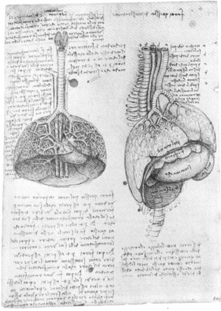
Although Leonardo is known to have dissected over 30 human cadavers, most of his drawings of the respiratory system are based on dissections of animals, including Figure 13.2 showing in beautiful detail the structure of the pig lung. In the commentary on this drawing, Leonardo considers the use of the ‘substance’ of the lung and extends Galen’s protective function of the lung parenchyma when he states that ‘the substance is interposed between these ramifications [of the trachea] and the ribs of the chest to act as a soft covering’. Structures entering the chest cavity are labelled a–e and their functions described:
He did however challenge some Galenic ideas by application of his engineering expertise. For example, he did not accept that the heart generated innate heat, instead writing that heat generation in the heart resulted from mechanical friction between the blood and the walls of the heart.4 Similarly, his engineering knowledge made him intrigued by the actions of the chest wall and respiratory muscles, including the complexities of defining the different function of internal and external intercostal muscles. For the diaphragm, Leonardo described four functions – dilating the lung for inspiration; pressing the stomach to drive food into the intestine; contracting with the abdominal muscles to drive out abdominal superfluities; and to separate the spiritual (thoracic) organs from the natural (abdominal) ones. Finally, he considered in detail the movements of the trachea and bronchi on breathing, showing them to dilate and open wider at branches on inspiration as shown to the right of Figure 13.3.
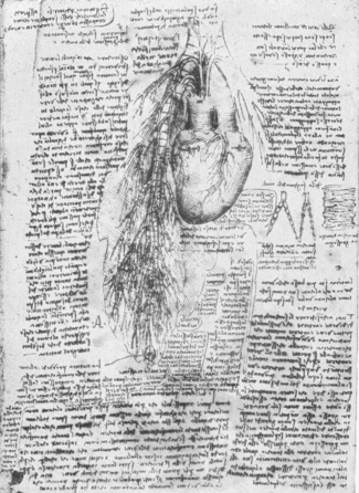
Fig. 13.3 Leonardo’s drawing of the pulmonary circulation in relation to the bronchi (c. 1513). Pulmonary vessels arise from several parts of the heart, leading Leonardo to propose a dual blood supply to the lung. Coronary arteries and veins can be clearly seen on the heart. At the lower end of the main drawing, Leonardo has drawn a small circle containing the letter N. The notes describe the structure as having ‘a crust, like a nutshell’ containing a ‘dust and watery humour’ possibly representing his discovery of a lung cyst16 or a tuberculous cavity.14
(The Royal Collection © 2010, Her Majesty Queen Elizabeth II.)
Leonardo and the bronchial circulation. In Figure 13.3, Leonardo depicts in detail the relationship of the pulmonary circulation to a bronchus. Much of the commentary in the drawing is concerned with the superiority of drawings rather than words to describe such anatomical configurations. The figure clearly shows a dual blood supply to the lung, and suggests that the smaller of these two supplies is to ‘nourish and vivify the trachea’. From this drawing, many writers have credited Leonardo with discovering the bronchial circulation, though this claim is disputed.16,17 The drawing is believed to be based on an ox, a species recently shown to have distinct small pulmonary veins draining directly into the left atrium, which may be those found by Leonardo.
The possibility of artistic license in his drawings has caused disputes that will never be resolved, such as that of the bronchial circulation. For example, in Figure 13.2 the perfectly branching pattern of the bronchi on the lung surface is clearly not based on true observation of pig lungs. In Figure 13.3 of ox lungs, the right upper lobe bronchus that arises directly from the trachea in this species is absent. However, in spite of these misgivings regarding his drawings, Leonardo’s genius in combining art, science and engineering in the study of physiology is undisputed.
Anatomy in the Renaissance
After Leonardo, the pursuit of medical knowledge in the universities continued, with anatomy in particular aided by the continuing resurgence of dissection and vivisection. Andreas Vesalius (1514–1564) is primarily remembered as the founder of modern anatomy, his dissections culminating in the publication in 1543 of De Humanis Corporis Fabrica, a book of seven volumes including over 250 anatomical illustrations (Figure 13.4).18 His ideas met with resistance from his contemporaries whenever his views were at odds with those of Galen, and this eventually forced Vesalius to cease his study of anatomy and to return to work as a physician. Nevertheless, the Fabrica continued to gain acceptance and became the foundation for future anatomy texts. Vesalius was also a skilled physiologist. He was the first to describe an experiment reproduced much later, in which a section of the chest wall of an animal was carefully removed without breaching the pleura beneath, so enabling direct observation of lung movements through the transparent pleura.6
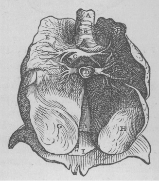
Fig. 13.4 Figure from Book VI of Vesalius’s Fabrica,18 showing an anterior view of the lungs after removal of the heart. A, oesophagus; B, trachea; C, pulmonary artery; D, pulmonary vein; I, diaphragm. E–H refer to the lobes of the lung – Vesalius’s illustrations always showed each lung to have four lobes.
(Reproduced by permission of the Special Collections, Leeds University Library.)
The pulmonary circulation.19 Unaware of the earlier writings of Ibn Al Nafis, Servetus (1509–1553), in his religious treatise Christianismi restitutio,20 again challenged the existence of Galen’s interventricular pores. He wrote that rather than passing through the middle wall of the heart, ‘blood is urged forward by a long course through the lung … and is poured from the pulmonary artery to the pulmonary vein’.21
Stay updated, free articles. Join our Telegram channel

Full access? Get Clinical Tree


