The Heart and Auscultation
PRETEST 
1. Where is the heart located in the chest, and what are the heart’s dimensions in the average adult?
2. What are systole and diastole?
3. How do electrical impulses stimulate the heart muscle to contract?
4. Name the precordial sites recommended for auscultation of heart sounds.
5. Which sound frequency is transmitted best by (a) the bell of the stethoscope and (b) the diaphragm of the stethoscope?
ANATOMY AND PHYSIOLOGY
Heart location and size
The heart is a powerful organ. Its primary functions are to pump deoxygenated blood to the lungs, where carbon dioxide-oxygen (CO2-O2) exchange takes place, and to pump oxygenated blood throughout the body. The top, or superior, part of the heart is called the base; the inferior, lateral part of the heart is called the apex. The average-sized adult heart is about 4 ¾″ (12 cm) long from the base to the apex and about 31/8 (8 cm) wide at its widest point. The heart gradually increases in size from infancy through early adulthood, and usually is slightly larger in males than in females.
Right and left sides
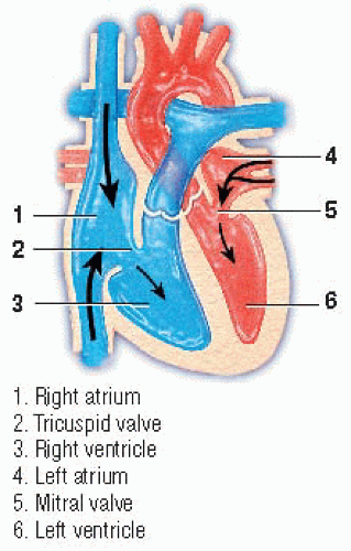
Anatomically, the heart is divided into two functional sides: the right side and the left side, which are separated by the septum. Each side is further divided into chambers. The right atrium and right ventricle are the right-sided chambers; the left atrium and left ventricle, the left-sided chambers.
Each chamber has unidirectional valves that separate the chambers and control blood flow. The right atrioventricular (AV), or tricuspid, valve separates the right atrium and right ventricle; the left AV, or mitral, valve separates the left atrium and left ventricle. The semilunar pulmonic valve separates the right ventricle and pulmonary artery, and the semilunar aortic valve separates the left ventricle and aorta.
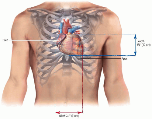 Dimensions of average-sized adult heart |
Blood flow
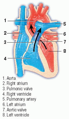
Deoxygenated, or venous, blood from nearly every tissue of the body flows into the right atrium via the superior and inferior vena cava. Venous blood from the heart tissue drains via the coronary sinus directly into the right atrium. The deoxygenated blood collects there until the tricuspid valve opens, and then it flows into the right ventricle. From the ventricle, it flows through the open pulmonic valve into the pulmonary artery, which distributes the deoxygenated blood to both lungs.
The complex process of CO2-O2 exchange occurs in the alveoli. Then the oxygenated blood flows back to the heart via the pulmonary veins and collects in the left atrium. From there it flows through the opened mitral valve into the left ventricle. When the aortic valve opens, the oxygenated blood flows into the aorta, which transports it to all body tissues.
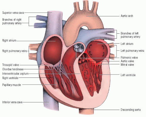 Internal structures of the heart |
 Systole |
Heart as a pump
A healthy heart in a 154-lb (70-kg) adult pumps about 5/8 gal (6 L) of blood per minute. Because the total blood volume in a healthy adult is about 13/8 gal (5.2 L), the heart pumps the total blood volume of the body in less than a minute. It performs this task continuously throughout an individual’s life.
An active process
The flow of deoxygenated and oxygenated blood through the heart, lungs, and rest of the body is an active process. The heart must generate enough pressure to pump blood through the arterial circulation. It does this by rhythmically contracting (systole) and relaxing (diastole).
During systole, heart muscle contraction raises the intracardiac pressures enough to systematically open the semilunar valves and pump a certain volume of blood. During diastole, the heart muscle relaxes, allowing the chambers to fill with blood again. This entire sequence is called the cardiac cycle.
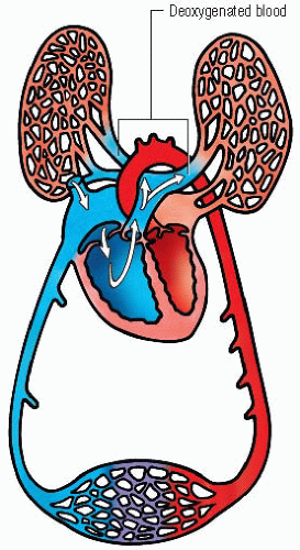 Pathway of deoxygenated blood through right heart to lungs |
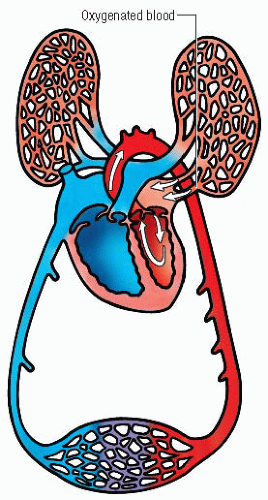 Pathway of oxygenated blood from lungs through left heart |
 Diastole |
The amount of blood pumped into the aorta each minute (cardiac output) depends largely on stroke volume. Stroke volume is the amount of blood pumped during each ventricular contraction.
As a person without heart disease ages, his heart usually becomes slightly larger and loses its contractile strength and efficiency. By age 70, cardiac output at rest has usually diminished by 30% to 35%.
Conduction system
Electrical impulses are primarily responsible for the rhythmic pumping action of the heart. These impulses are discharged automatically from specialized cardiac cells that stimulate the heart muscle to contract. The specialized cardiac cells and fibers within the conduction system work together to produce a contraction. The impulses fired from the sinoatrial (SA) node, located just below the entrance of the superior vena cava in the right atrium, normally initiate the cardiac cycle, and they usually travel along specific pathways.
After the impulse
When the electrical impulse leaves the SA node, it travels along the atrial conduction pathways located in the atrial walls, initiating atrial systole (or atrial contraction). The impulse rapidly reaches the AV node, located near the AV junction. From the AV node,
the impulse travels to the bundle of His located in the interventricular septum. Here, the bundle of His divides into the right and left bundle branches. These branches further divide into the tiny Purkinje fibers located throughout the ventricular walls. As the impulse travels through the bundle branches and Purkinje fibers, it initiates ventricular systole or ventricular contraction.
the impulse travels to the bundle of His located in the interventricular septum. Here, the bundle of His divides into the right and left bundle branches. These branches further divide into the tiny Purkinje fibers located throughout the ventricular walls. As the impulse travels through the bundle branches and Purkinje fibers, it initiates ventricular systole or ventricular contraction.
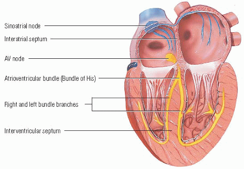 Cardiac conduction |
As the myocardium of the aging heart becomes more irritable, extrasystoles may occur, along with sinus arrhythmias and bradycardias. In addition, increased fibrous tissue infiltrates the SA node and internodal tracts, possibly causing atrial fibrillation and flutter.
Cardiac cycle
Several mechanical events occurring simultaneously during the cardiac cycle enable the heart to pump oxygenated blood throughout the body.
During atrial diastole, when the right atrium and left atrium are relaxed, blood flows into the atria. When the pressure in the atria exceeds the pressure in the ventricles, the tricuspid and mitral valves open, allowing blood to flow rapidly into the relaxed ventricles. The SA node, the pacemaker in a normally functioning heart, initiates an electrical impulse. The impulse is conducted via internodal pathways to both atria and results in atrial contraction. This contraction—atrial systole—pushes more blood from the atria to complete ventricular filling. The impulse is slowly conducted through the AV node, which allows time for the atria to contract completely and fill the ventricles with blood. From the
AV node, the impulse is rapidly conducted through the bundle of His and its branches. The Purkinje fibers receive the impulse and directly stimulate the ventricular myocardium. A forceful contraction of both ventricles opens the pulmonic and aortic valves and ejects blood into the pulmonary artery and aorta.
AV node, the impulse is rapidly conducted through the bundle of His and its branches. The Purkinje fibers receive the impulse and directly stimulate the ventricular myocardium. A forceful contraction of both ventricles opens the pulmonic and aortic valves and ejects blood into the pulmonary artery and aorta.
Stay updated, free articles. Join our Telegram channel

Full access? Get Clinical Tree


