(1)
Anatomy Department, University of Medicine and Pharmacy Carol Davila, Bucharest, Romania
Abstract
During this period, the developing heart goes through several developing phases; they shall be named according to each dominant stage.
1.1 The General Development Period [1, 2]
During this period, the developing heart goes through several developing phases; they shall be named according to each dominant stage.
1.1.1 The Germ Disk Stage
In the 18th and 19th day, the developing embryo is found in the trilaminar germ disk stage, with the following composing part:
The ectodermal tissue that exhibits the primitive streak on its dorsal surface (a central sulcus lying longitudinally) along side which the right to left symmetry is orientated.
the mesodermal tissue
the endodermal tissue
The trilaminar germ disk is flanked by the amniotic cavity and the yolk sac.
1.1.2 The Formation of the Endocardial Tubes
(A)
The differentiation of the cardiogenic area (the cardiogenic mesoderm) is created as a meshing of the mesoderm into the cephalic end of the germ disk with the mesoderm located above the prechordal plate (the future oropharyngeal membrane)
(B)
The formation of the two tubes takes place as follows:
The cardiogenic mesoderm wraps around the upper end of the neural plate – as a right and left cardiogenic mesodermal plane (the primary cardiogenic fields) [3]. A transversal section performed here shows that groups of cardiogenic angioblastic cells are found along each side of the lateral mesoderm of the germ disk. These two cellular groups form two full cords which immediately tunnel creating the right and left endocardial tubes.
In the germ disk there are multiple movements of the various cellular groups. The developing embryo undergoes folding movements in a transversal and longitudinal direction while other cells undergo the so called process of descensus.
The embryonic movements are not self initiated but they result from the cellular multiplication, differentiation and variable growth rates of the neighboring regions.
1.1.3 The Primitive Heart Tube Formation
This formation process occurs on the 22nd day, by the median merging of the two tubes and the transversal folding of the embryonic disk.
As the embryonic transversal folding occurs, the two endocardial tubes move closer. A fusing process follows that creates the primitive heart tube found in the ventral mesentery of the primitive gut.
This tube has a dorsal connection with the anterior portion of the primitive gut and is attached to the walls of the future pericardial cavity by groups of cells that form the dorsal mesocardium, the ventral mesocardium (this structure quickly disappears), the arterial and venous mesocardium (Fig. 1.1).
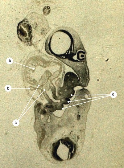
Fig. 1.1
Arterial mesocardium represented by the truncus arteriosus and branchial arches region junction. a Primitive ventricle, b right atrium, c endocardial crests in the truncus arteriosus, d branchial arches region
The endocardium develops from the primitive cardiac tube. The adjacent mesoderm called the mioepicardic plate, gives rise to the developing myocardium and epicardium. Between the endocardium and the plate there is a new structure called the cardiac jelly which is invaded by endocardiac cells and leads to the formation of the cardioglia (Figs. 1.2 and 1.3).
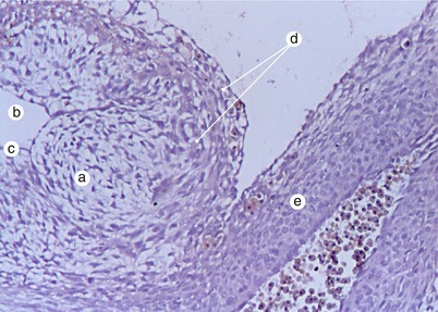
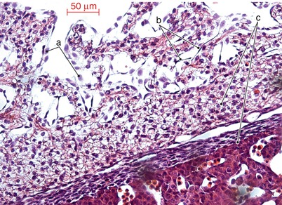
Fig. 1.2
Myoepicardial plate and cardioglia. a Cardioglia, b ventricle, c endocardium, d myoepicardial plate, e truncus arteriosus
Fig. 1.3
Myoepicardial plate and endocardium. a Blood cells in the ventricular cavity organized by the contact with the endocardium, b endocardium, c myoepicardial plate
The cardiac jelly contains about 50 proteins, such as: collagen, fibronectin, fibrillin, fibulin and growth factors [4].
1.1.4 The Formation of the Cardiac Prominence
The cardiac prominence becomes structured in the following way: the transversal folding movement occurs simultaneously with the longitudinal folding of the germ disk. The primitive cardiac tube location lies on the ventral side of the embryonic body, inferior to the stomodeum in the future cervical region. The cells proliferate and differentiate, creating the endocardial prominence. This prominence lies superiorly to the transverse septum and the vitelline sac. A more correct terminology is the hepatocardiac prominence (Fig. 1.4)
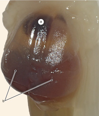
Fig. 1.4
Hepatocardiac bulge. a Cardiac bulge, b hepatic bulge
Between the heart and hepatic primordium lies the transverse septum, a large area of “embryologic unrest” which influences the hepatic and cardiac development.
Throughout the development stage, the heart undergoes a process of descensus. More precisely, the heart descends from the cervical to the thoracic area, above the diaphragm (already differentiated from the septum transversum). This process is not considered a cardiac descending as much as a growth at different speeds of the neighboring regions (pseudodescensus). On the 23rd day the first inefficient (arrhythmic) contractions occur with progression to the 28th day when they become rhythmic and efficient.
1.1.5 The Stage of Linear Cardiac Tube
As a result of the different types of movements and due to the disappearance of the ventral mesocardium, the cardiac tube remains fixed at the extremities, superior through the arterial mesocardium and inferior through the venous mesocardium (including the sinus venosus). The sinus is fixed to the transverse septum. Actually the horns of the sinus arise from the transverse septum and connect above it. The mesocardial sinus covers the space between the two horns and continues posteriorly with the dorsal mesocardium, tying the atrium and sinus venosus to the primitive gut (Figs. 1.5 and 1.6).
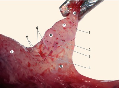
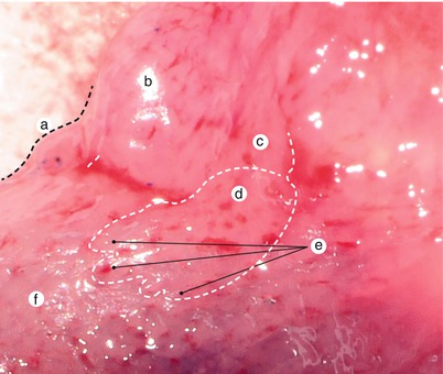
Fig. 1.5
Primitive cardiac tube with dorsal mesocardium – lateral view. a Dorsal aortae, b aortic bulb, c primitive bulb, d primitive ventricle, e primitive atrium, f septum transversum, g dorsal mesocardium undergoing reduction, 1 bulbo-aortic sulcus, 2 interbulbar sulcus, 3 bulboventricular sulcus, 4 atrioventricular sulcus
Fig. 1.6
Septum transversum with sinus horns, detail. Left anterolateral view. a Right primitive ventricle, b primitive bulbus, c left primitive ventricle, d left primitive atrium, e sinus tributaries, f septum transversum
Immediately superior to the sinus venosus, the walls of the primitive atrium reflect posteriorly, between the leaflets of the dorsal mesocardium, creating the atrio (myocardial)- mesodermal junction. The junction is therefore created between the cardiac mesoderm and the mesoderm lying anterior to the primitive gut [5] (Fig. 1.7)
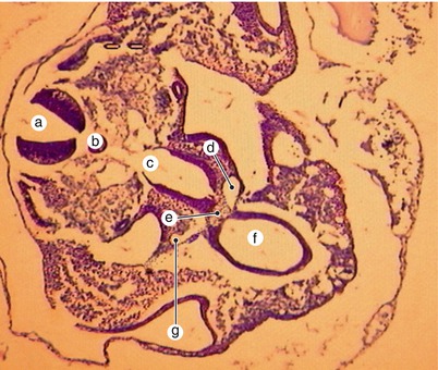
Fig. 1.7
Transverse section through the primitive cardiac tube at the level of the sinoatrial junction. a Neural tube, b notochord, c primitive intestine, d left sinusal horn, e dorsal mesocardium taenia, f endocardial cardiac tube, g right sinusal horn
This junction is a true mesodermal pedicle of the primitive atrium, covered on its exterior by the dorsal mesocardium.
Stay updated, free articles. Join our Telegram channel

Full access? Get Clinical Tree


