(1)
Ospedale Civile di Vigevano, Vigevano, Italy
Introduction
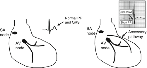
In its most common form, the Wolff-Parkinson-White (WPW) syndrome is caused by the presence of an embryonic remnant consisting of an accessory atrioventricular pathway. When the pathway bypasses the AV node and connects the atria directly to the ventricles, it is referred to as the bundle of Kent. Less frequently, the anomalous connection is between the AV node or bundle of His and the ventricles, and it is referred to as the bundle of Mahaim. The bundle of Kent is by far the more common variant. In this case, the excitation wavefront travels simultaneously through the usual pathway (the AV node, which normally slows conduction) and much more rapidly through the accessory pathway, thereby activating the ventricles earlier than usual. As a result, the P-R interval is shortened, and the initial part of the ventricular complex is slurred or notched. This distortion is referred to as a delta wave, and it is pathognomonic for the WPW syndrome. The QRS complex in this case is actually a fusion of the complexes resulting from the two pathways of ventricular activation (Figs. 7.1 and 7.2).


Fig. 7.1
Right: Diagram of the Kent bundle-type accessory conduction pathway and typical ECG showing a delta wave. Left: Diagram pf the normal excitation-conduction system and normal ECG

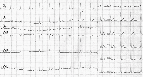
Fig. 7.2
ECG showing left ventricular preexcitation: sinus rhythm (rate 70 bpm), short PR interval (0.08 s), QRS duration 0.12 s. The delta wave is positive in the precordial leads and in aVF with an intermediate electrical axis in the frontal plane
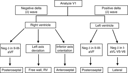
The accessory pathway may be located in the free wall of the left or right ventricle or in the interventricular septum. In some cases, there may be more than one. In around 15 % of cases, it conducts exclusively in a retrograde direction and is referred to as a concealed accessory pathway. Preexcitation has been historically classified as type A or type B (although this distinction of limited value). Accessory pathways located in the left side of the heart give rise to type A preexcitation, with premature activation of the left ventricle. The QRS complexes in this case are generally positive in the right precordial leads and exhibit a right bundle-branch-block (RBBB) pattern. Right-heart pathways produce type B preexcitation, with premature activation of the right ventricle and QRS complexes with a left bundle-branch-block (LBBB) morphology. When type-B pathways are located posteriorly, they often give rise to Q waves or QS complexes in leads II, III, and aVF, findings that can be misinterpreted as signs of an inferior myocardial infarction. The exact location of the accessory pathway cannot always be determined on the basis of surface electrocardiographic findings. Diagnostic algorithms have been developed for this purpose although their sensitivity and specificity varies (Fig. 7.3). In general, the delta-wave vector is directed away from the ventricular zone that is pre-excited by the accessory pathway (Table 7.1).


