(1)
Ospedale Civile di Vigevano, Vigevano, Italy
Acute Cor Pulmonale—Pulmonary Embolism
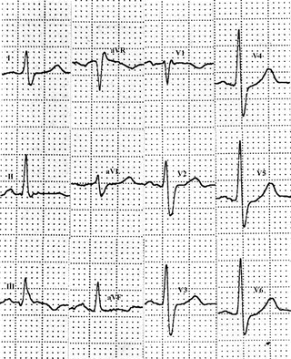
The electrocardiogram (ECG) is a specific but relatively insensitive tool for diagnosing acute cor pulmonale, which is almost always caused by pulmonary embolism. Acute overload of the right ventricle is manifested by a typical triad of ECG features referred to as the S1Q3T3 complex or McGinn and White triad. It includes the presence of S waves in lead I and Q waves with T-wave inversion in lead III (Fig. 14.1). Acute cor pulmonale can also be associated with right axis deviation, transient right bundle-branch block (RBBB), and T-wave inversion in the right precordial leads (Fig. 14.2). It has been suggested that inverted T waves in lead V2 or V3 are a common ECG sign of massive pulmonary embolism. In another study on pulmonary embolism, the pseudoinfarction pattern (QR in V1) and T-wave inversion in lead V2 were strongly correlated with the presence of right ventricular dysfunction and independent predictors of an unfavorable clinical outcome.


Fig. 14.1


Massive pulmonary embolism: The ECG shows the classic S1Q3T3 pattern (McGinn-White triad) and incomplete RBBB
Stay updated, free articles. Join our Telegram channel

Full access? Get Clinical Tree


