(1)
Ospedale Civile di Vigevano, Vigevano, Italy
Acute Pericarditis
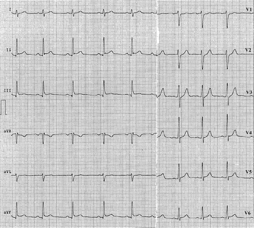
Acute pericarditis is associated with inflammation of the subepicardial layers of the myocardium, which are in contact with the pericardium. The electrocardiogram (ECG) can be useful in the diagnosis of this disorder, revealing diffuse ST-segment and T-wave changes that evolve in a characteristic manner and are phase-specific.
Within a few hours of symptom onset, ST-segment elevation appears. It reflects abnormal repolarization secondary to pericardial inflammation and is the most sensitive ECG criterion for pericarditis. The ST-segment elevation is diffuse and concordant with the T waves. Its magnitude (the amplitude rarely exceeds 5 mm) and upwardly concave shape distinguish it from the convex ST-segment elevation associated with transmural ischemia. The ST-segment alterations associated with acute pericarditis are generalized because the pericardial inflammation is usually diffuse. ST-segment elevation is never observed in lead aVR, but ST-segment depression may well be present in lead aVR or V1 (Fig. 13.1). Acute pericarditis can also be distinguished from acute myocardial infarction by the absence of reciprocal ST-segment depression in leads opposite to those displaying elevation and by the invariable absence of necrosis-related Q waves (excluding those reflecting a prior infarction). During this phase, the presence of a significant pericardial effusion leads to diffuse reductions in the amplitudes of all electrocardiographic waves owing to the electrical insulating effects of the large fluid collection (Fig. 13.2). In subsequent phases, the ST-segment elevation diminishes, and T-wave inversion appears. It can persist for long periods of time but disappears as the clinical picture improves (Fig. 13.3).


