6
The ECG in Patients with Breathlessness
The ECG in disorders affecting the left side of the heart
The ECG in left atrial hypertrophy
The ECG in left ventricular hypertrophy
ECGs that can mimic left ventricular hypertrophy
The ECG in disorders affecting the right side of the heart
The ECG in right atrial hypertrophy
The ECG in right ventricular hypertrophy
HISTORY AND EXAMINATION
There are many causes of breathlessness (see Box 6.1). Everyone is breathless at times, but people who are physically unfit or who are overweight will be more breathless than others. Breathlessness can also result from anxiety, but when it is due to physical illness the important causes are anaemia, heart disease and lung disease; a combination of causes is common. Apart from ischaemia (see Ch. 5), cardiac diseases causing breathlessness include valve disease, cardiomyopathy, and myocarditis including acute rheumatic fever. Boxes 6.2–6.6 summarize the effects of these conditions on the heart, and the corresponding ECG features. The most important function of the history is to help to determine whether the patient does indeed have a physical illness and, if so, which system is affected.
Breathlessness in heart disease is due to either increased lung stiffness, as a result of pulmonary congestion, or pulmonary oedema. Pulmonary congestion occurs when the left atrial pressure is high. A high left atrial pressure occurs either in mitral stenosis or in left ventricular failure. Pulmonary oedema occurs when the left atrial pressure exceeds the oncotic pressure exerted by the plasma proteins.
RHYTHM PROBLEMS
A sudden rhythm change is a common cause of breathlessness, and even of frank pulmonary oedema. Arrhythmias can be paroxysmal, so the patient may be in sinus rhythm when examined, and a patient who is suddenly breathless may not be aware of an arrhythmia. When sudden breathlessness is associated with palpitations it is important to establish whether the breathlessness or the palpitations came first palpitations following breathlessness may be due to the sinus tachycardia of anxiety. The ECG in Figure 6.1 is from a patient who developed pulmonary oedema due to the onset of uncontrolled atrial fibrillation.
Less dramatic rhythm abnormalities can also contribute to breathlessness, especially to breathlessness on exertion. This is true of both fast and slow rhythms. The ECG in Figure 6.2 is from a patient who had atrial fibrillation but who was breathless on exercise partly because of coupled ventricular extrasystoles, which markedly reduced cardiac output as a result of an effective halving of the heart rate.
THE ECG IN DISORDERS AFFECTING THE LEFT SIDE OF THE HEART
THE ECG IN LEFT ATRIAL HYPERTROPHY
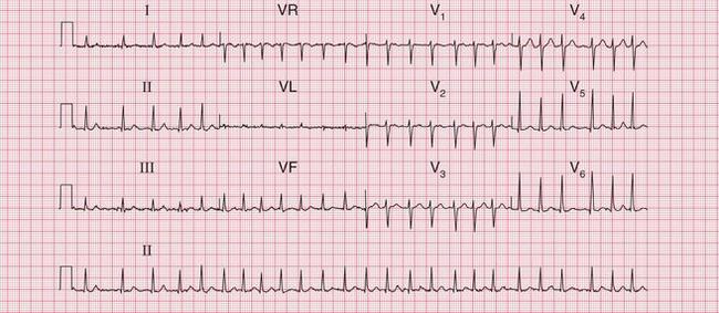
Left atrial hypertrophy causes a double (bifid) P wave. Left atrial hypertrophy without left ventricular hypertrophy is classically due to mitral stenosis, so the bifid P wave is sometimes called ‘P mitrale’. This is misleading, because most patients whose ECGs have bifid P waves either have left ventricular hypertrophy that is not obvious on the ECG or and perhaps this is more common have a perfectly normal heart. The bifid P wave is thus not a useful measure of left atrial hypertrophy.
Figure 6.3 shows an ECG with a bifid P wave indicating left atrial hypertrophy. This was confirmed by echocardiography in the patient, who also had concentric left ventricular hypertrophy due to hypertension.
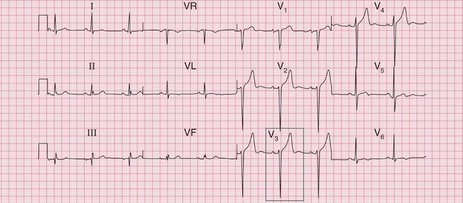
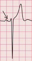
Fig. 6.3 Left atrial hypertrophy and left ventricular hypertrophy
Note
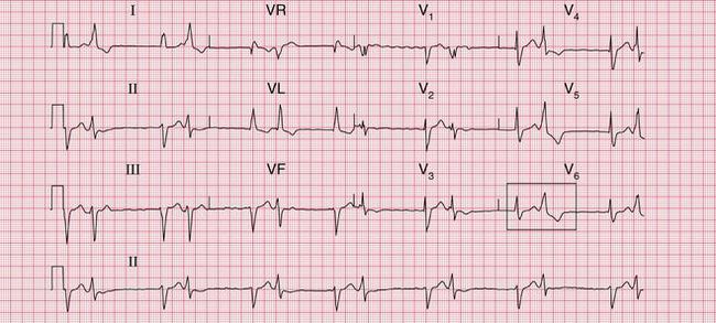
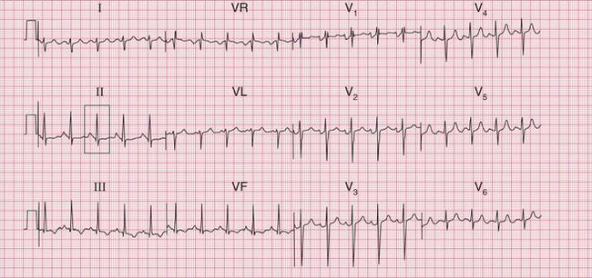
Significant mitral stenosis usually but not always leads to atrial fibrillation, in which no P waves, bifid or otherwise, can be seen. Occasional patients, such as the one whose ECG is shown in Figure 6.4, develop pulmonary hypertension and remain in sinus rhythm. There is then a combination of a bifid P wave with evidence of right ventricular hypertrophy. This combination does allow a confident diagnosis of severe mitral stenosis.
THE ECG IN LEFT VENTRICULAR HYPERTROPHY
Left ventricular hypertrophy may be caused by hypertension, aortic stenosis or incompetence, or mitral incompetence.
The ECG features of left ventricular hypertrophy are:
• an increased height of the QRS comple
• inverted T waves in the leads that ‘look at’ the left ventricle I, VL and V5-V6.
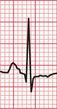
Unfortunately, voltage criteria have a low sensitivity as detectors of left ventricular hypertrophy, and are essentially useless. They would frequently lead to a diagnosis of left ventricular hypertrophy in perfectly healthy young men, even in those who are not athletic ( Fig. 6.5).
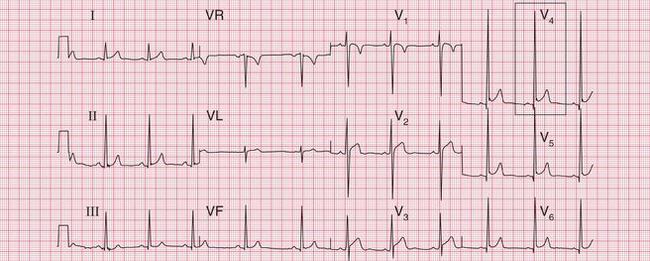
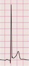
Fig. 6.5 Probably normal ECG
Note
< div class='tao-gold-member'>
Stay updated, free articles. Join our Telegram channel

Full access? Get Clinical Tree


