5
The ECG in Healthy Subjects
The ECG is frequently used in ‘health screening’, but it is important to remember that not all those who are screened are really asymptomatic – the procedure may be used as an alternative to seeking medical advice. On the other hand, people being screened may be totally free of symptoms and yet important abnormalities may be evident from their ECGs. For example, Figure 5.1 shows the ECG of an asymptomatic patient which, quite unexpectedly, revealed atrial fibrillation. Abnormalities are uncommon in this group of individuals, but perhaps are the best reason for using the ECG in screening. All the ECGs in this chapter came from health screening clinics, and we will assume that the individuals considered themselves to be healthy.
THE NORMAL CARDIAC RHYTHM
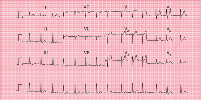
Sinus rhythm is the only truly normal rhythm. ‘Sinus bradycardia’ is sometimes said to be present when the heart rate is below 60/min, and ‘sinus tachycardia’ is sometimes used for heart rates above 100/min ( Box 5.1), but these terms are really not helpful, and it is far more useful to describe a patient as having ‘sinus rhythm at x/min’ ( Fig. 5.2).
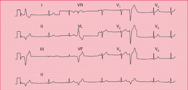
EXTRASYSTOLES
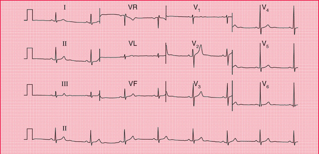
Supraventricular extrasystoles are of no clinical significance, although atrial extrasystoles need to be differentiated from the variations in beat- to-beat interval which occur in sinus rhythm ( Figs 5.3 and 5.4). Automated ECG reporting often fails to do this.
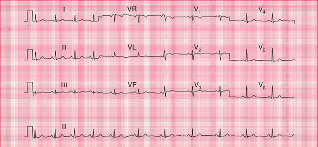
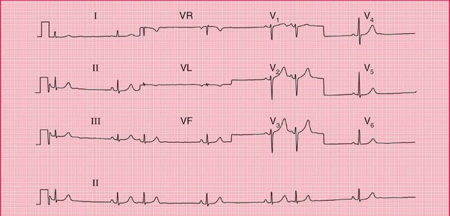
Fig 5.4 Atrial extrasystoles
Note
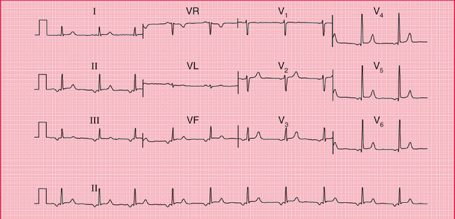
Occasional ventricular extrasystoles are experienced by many people with normal hearts. Frequent ventricular extrasystoles ( Fig. 5.5) may indicate heart disease, and in a large population their presence does identify a group with a higher than average risk of developing cardiac problems. In an individual patient, however, their presence is not a good predictor of such risk.
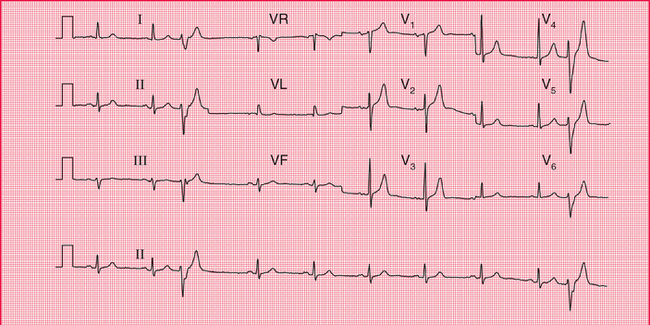
ECTOPIC ATRIAL RHYTHM
When depolarization is initiated from a focus in the atrium rather than in the sinoatrial node, an ‘ectopic atrial rhythm’ is present ( Fig. 5.6). This does not cause symptoms and is usually of no clinical significance. It is not an uncommon finding in individuals being screened.
THE P WAVE
Tall P waves may be due to right atrial hypertrophy, and are significant if there is evidence of right ventricular hypertrophy as well. Tall P waves alone may indicate tricuspid stenosis, but this is rare. If the patient is well and has no abnormal physical signs, ‘tall’ P waves are probably within the normal limits.
 For an example mitral stenosis, see p. 293
Bifid P waves in the absence of signs of associated left ventricular hypertrophy can indicate mitral stenosis (now fairly rare), but a bifid and not particularly prolonged P wave is often seen in the anterior leads of normal ECGs. Figure 5.7 shows the ECG of an asymptomatic patient with a clinically normal heart.
The P waves of atrial extrasystoles tend to be abnormally shaped compared to the P waves of the sinus beats of the same patient ( Fig. 5.4).
 For more on hyperkalaemia, see p. 331
CONDUCTION
The ECG in Figure 5.8 was recorded from a healthy, asymptomatic individual at a screening examination. Nevertheless, PR interval prolongation to this extent is probably evidence of disease of the conducting tissue.
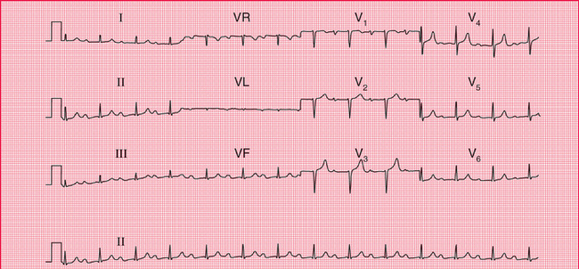
Fig 5.8 First degree heart block
Note
< div class='tao-gold-member'>
Stay updated, free articles. Join our Telegram channel

Full access? Get Clinical Tree


