Fig. 16.1
Admission ECG demonstrating widespread ST segment changes
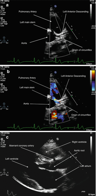
Echocardiogram demonstrated mild left ventricular hypertrophy (interventricular septum in diastole 1.7 cm, posterior wall 1.2 cm). Inspection of his coronary arteries demonstrated a left coronary artery arising aberrantly from the right coronary sinus and coursing between the aorta and pulmonary artery (Fig. 16.2a–c). There were no regional wall motion abnormalities and function was normal with a fractional shortening of 37 %.


Fig. 16.2
(a) Echocardiographic 2D image of left coronary artery demonstrating interarterial segment of LCA followed by normal bifurcation. (b) Colour flow image of LCA demonstrating interarterial segment. (c) 2D image in the parasternal long axis showing the aberrant coronary artery
A computed tomogram (CT) of his chest was performed to further assess coronary anatomy (Fig. 16.3a, b). This demonstrated the left main stem arising from the right coronary sinus separate from the right coronary artery with a hooded orifice and narrowing through its inter-arterial portion with no intramural course. Cardiac function was normal.


Fig. 16.3
(a) Transverse slice from CT demonstrating left main coronary artery arising from anterior (right coronary) sinus and coursing between aorta and pulmonary artery. (b) CT reconstruction of aortic root. The aorta is coloured red and the pulmonary artery grey. The left coronary is seen emerging acutely from the right sinus and then coursing between the great vessels
He was discussed at the multidisciplinary meeting and the decision made for surgical repair. At surgery, he was found to have a slit-like orifice of his left main stem from the right aortic sinus with an inter-arterial course. His left coronary artery was reimplanted into the left sinus. He came off bypass easily and made an uneventful recovery.
Follow-up investigations included an exercise tolerance test and cardiac magnetic resonance imaging. On exercise testing he has completed 12 min of the Bruce protocol with no ischaemic changes. MRI scan has shown an unobstructed course of the left coronary artery and good left ventricular function with no evidence of myocardial scarring. He has remained perfectly well in the 3 years since his operation.
Discussion
Normal Coronary Anatomy
The right coronary artery normally arises from the right coronary sinus, before coursing between the right atrial appendage and right ventricular infundibulum through the right atrioventricular groove. A high conal branch emerges in 50 % followed by a sinoatrial nodal branch in 50 %. Marginal vessels then emerge to supply the right ventricular free wall. The vessel then continues posteriorly in the right atrioventricular groove to the posterior interventricular groove, where in 90 %, the posterior descending coronary artery emerges to run in the posterior AV groove and supply the posterior third of the interventricular septum.
The left coronary artery (LCA) emerges from the left coronary sinus and covers a short distance under the orifice of the left atrial appendage before dividing into the left circumflex (LCx) and left anterior descending (LAD). The LCx runs in the left atrioventricular groove, giving off obtuse marginal branches which supply the free wall of the left ventricle. In the majority of patients, the circumflex ends towards the posterior interventricular groove, although in 10 % the vessel continues into the posterior interventricular groove as the posterior descending artery. The LAD passes down the anterior interventricular groove, giving a left conal branch, diagonal branches to left and right ventricular free walls and septal perforator vessels to feed the anterior two-thirds of the interventricular septum.
The atrioventricular nodal artery is given by the right coronary artery in 50 %, and in the remainder from the left coronary. A similar division of frequencies is seen for the origin of the sinoatrial artery.
< div class='tao-gold-member'>
Only gold members can continue reading. Log In or Register to continue
Stay updated, free articles. Join our Telegram channel

Full access? Get Clinical Tree


