The Anatomy of the Heart’s Electrical System
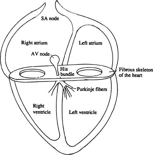
The heart’s electrical impulse originates in the sinoatrial (SA) node, located high in the right atrium near the superior vena cava. The impulse leaves the SA node and spreads radially across both atria. When the impulse reaches the atrioventricular (AV) groove, it encounters the “skeleton of the heart,” the fibrous structure to which the valve rings are attached, and which separates the atria from the ventricles. This fibrous structure is electrically inert and acts as an insulator—the electrical impulse cannot cross this structure. Thus the electrical impulse would be prevented from crossing over to the ventricular side of the AV groove if not for the specialized AV conducting tissues: the AV node and the bundle of His (Figure 1.1).
As the electrical impulse enters the AV node, its conduction is slowed because of the electrophysiologic properties of the AV nodal tissue. This slowing is reflected in the PR interval on the surface electrocardiogram (ECG). Leaving the AV node, the electrical impulse enters the His bundle, the most proximal part of the rapidly conducting His–Purkinje system. The His bundle penetrates the fibrous skeleton and delivers the impulse to the ventricular side of the AV groove.
Once on the ventricular side, the electrical impulse follows the His bundle as it branches into the right and left bundle branches. Branching of the Purkinje fibers continues distally to the furthermost reaches of the ventricular myocardium. The electrical impulse is thus rapidly distributed throughout the ventricles.
Hence, the heart’s electrical system is designed to organize the sequence of myocardial contraction with each heartbeat. As the electrical impulse spreads over the atria toward the AV groove, the atria contract. The delay provided by the AV node allows for complete atrial emptying before the electrical impulse reaches the ventricles. Once the impulse leaves the AV node, it is distributed rapidly throughout the ventricular muscle by the Purkinje fibers, providing for brisk and orderly ventricular contraction.
We next consider the character of the electrical impulse, its generation, and its propagation.
The Cardiac Action Potential
The cardiac action potential is one of the most despised and misunderstood topics in cardiology. The fact that electrophysiologists claim to understand it is also a leading cause of the mystique that surrounds them and their favorite test, the electrophysiology study. Because the purpose of this book is to debunk the mystery of electrophysiology studies, we must confront the action potential and learn to understand it. Fortunately, this is far easier than legend would have it.
Although most of us would like to think of cardiac arrhythmias as an irritation or “itch” of the heart (and of antiarrhythmic drugs as a balm or a salve that soothes the itch), this conceptualization of arrhythmias is wrong and leads to the faulty management of patients with arrhythmias. In fact, the behavior of the heart’s electrical impulse and of the cardiac rhythm is largely determined by the shape of the action potential; the effect of antiarrhythmic drugs is determined by how they change that shape.
The inside of the cardiac cell, like all living cells, has a negative electrical charge compared to the outside of the cell. The resulting voltage difference across the cell membrane is called the transmembrane potential. The resting transmembrane potential (which is −80 to −90 mV in cardiac muscle) is the result of an accumulation of negatively charged molecules (called ions) within the cell. Most cells are happy with this arrangement and live out their lives without considering any other possibilities.
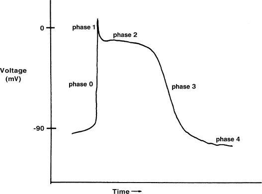
Cardiac cells, however, are excitable cells. When excitable cells are stimulated appropriately, tiny pores or channels in the cell membrane open and close sequentially in a stereotyped fashion. The opening of these channels allows ions to travel back and forth across the cell membrane (again in a stereotyped fashion), leading to patterned changes in the transmembrane potential. When these stereotypic voltage changes are graphed against time, the result is the cardiac action potential (Figure 1.2). The action potential is thus a reflection of the electrical activity of a single cardiac cell.
As can be seen in Figure 1.2, the action potential is classically divided into five phases. However, it is most helpful to consider the action potential in terms of three general phases: depolarization, repolarization, and the resting phase.
Depolarization
The depolarization phase (phase 0) is where the action of the action potential is. Depolarization occurs when the rapid sodium channels in the cell membrane are stimulated to open. When this happens, positively charged sodium ions rush into the cell, causing a rapid, positively directed change in the transmembrane potential. The resultant voltage spike is called depolarization. When we speak of the heart’s electrical impulse, we are speaking of this depolarization.
Depolarization of one cell tends to cause adjacent cardiac cells to depolarize, because the voltage spike of a cell’s depolarization causes the sodium channels in the nearby cells to open. Thus, once a cardiac cell is stimulated to depolarize, the wave of depolarization (the electrical impulse) is propagated across the heart, cell by cell.
Further, the speed of depolarization of a cell (reflected by the slope of phase 0 of the action potential) determines how soon the next cell will depolarize, and thus determines the speed at which the electrical impulse is propagated across the heart. If we do something to change the speed at which sodium ions enter the cell (and thus change the slope of phase 0), we therefore change the speed of conduction (the conduction velocity) of cardiac tissue.
Repolarization
Once a cell is depolarized, it cannot be depolarized again until the ionic fluxes that occur during depolarization are reversed. The process of getting the ions back to where they started is called repolarization. The repolarization of the cardiac cell roughly corresponds to phases 1 through 3 (i.e. the width) of the action potential. Because a second depolarization cannot take place until repolarization occurs, the time from the end of phase 0 to late in phase 3 is called the refractory period of cardiac tissue.
Repolarization of the cardiac cells is complex and poorly understood. Fortunately, the main ideas behind repolarization are simple: 1) repolarization returns the cardiac action potential to the resting transmembrane potential; 2) it takes time to do this; and 3) the time that it takes to do this, roughly corresponding to the width of the action potential, is the refractory period of cardiac tissue.
There is an additional point of interest regarding repolarization of the cardiac action potential. Phase 2 of the action potential, the so-called plateau phase, can be viewed as interrupting and prolonging the repolarization that begins in phase 1. This plateau phase, which is unique to cardiac cells (e.g. it is not seen in nerve cells), gives duration to the cardiac potential. It is mediated by the slow calcium channels, which allow positively charged calcium ions to slowly enter the cell, thus interrupting repolarization and prolonging the refractory period. The calcium channels have other important effects in electrophysiology, as we will see.
The Resting Phase
For most cardiac cells, the resting phase (the period of time between action potentials, corresponding to phase 4) is quiescent, and there is no net movement of ions across the cell membrane.
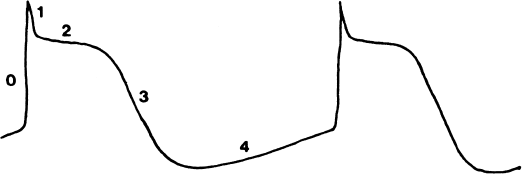
For some cells, however, the so-called resting phase is not quiescent. In these cells, there is leakage of ions back and forth across the cell membrane during phase 4, in such a way as to cause a gradual increase in transmembrane potential (Figure 1.3). When the transmembrane potential is high enough (i.e. when it reaches the threshold voltage), the appropriate channels are activated to cause the cell to depolarize. Because this depolarization, like any depolarization, can stimulate nearby cells to depolarize in turn, the spontaneously generated electrical impulse can be propagated across the heart. This phase 4 activity, which leads to spontaneous depolarization, is called automaticity.
Figure 1.3 Automaticity. In some cardiac cells, there is a leakage of ions across the cell membrane during phase 4, in such a way as to cause a gradual, positively directed change in transmembrane voltage. When the transmembrane voltage becomes sufficiently positive, the appropriate channels are activated to automatically generate another action potential. This spontaneous generation of action potentials due to phase 4 activity is called automaticity.

Stay updated, free articles. Join our Telegram channel

Full access? Get Clinical Tree


