Fig. 3.1
Non-video and video bronchoscopes
The true “video” bronchoscope (Fig. 3.1) did not become available until 1987 when the charged coupled device technology (CCD chip) was able to be miniaturized to a point that it could be used in the endoscope. This advancement allowed tremendous improvements in image quality as well as the ability to integrate the imaging into a monitor for others to view.
While the majority of bronchoscopes being produced today are true video scopes, there are still isolated instances where space limitations require fiber-optic bundles be used through most of the bronchoscope for image creation. This “hybrid” bronchoscope utilizes a combination of video and non-video technology to provide an improved image over a true fiber-optic system.
An example of this hybrid technology is the current bronchoscope with an ultrasound transducer built into the distal end used to view mediastinal lymph nodes (Fig. 3.2). Because of the size of the transducer, there is not enough space to also place a CCD chip for optimum imaging, so the insertion tube has a fiber-optic bundle used to transmit the image to a point in the control head where there is enough space to place a CCD chip. Until the technology is advanced to create a CCD chip small enough, fiber-optic technology is used to assist in providing imaging. Here is a good example of the development of a “new” type of bronchoscope which relies in part on the “old” non-video technology for its imaging.
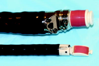

Fig. 3.2
Ultrasound endoscopes
The Size of the Bronchoscope
Since most bronchoscopes all perform basic functions allowing the operator to navigate the airways under direct vision and allow for sampling and video documentation, then the difference are the functions that the operator feels are needed to their specific patient population. Probably the biggest factor to consider is the size of the scope you may need. There are two factors to consider here: one is the outer diameter of the scope (insertion tube) and the other is the size of the working channel (the channel that accessories are placed for either sampling or treating). One would assume that there should be a direct relationship between the two, but that is not always the case. Sometimes, a scope will have to compromise the size of the working channel to allow for another technology to use some of that space. There are two examples that come to mind. One is the case of the dedicated ultrasound bronchoscope. The outer diameter of the scope is as large as one could probably make a scope to fit through the vocal cords of an adult, yet the working channel is that of only a standard scope (2 mm). That is because the ultrasound transducer has to take up much of the extra space in the insertion tube. The other is the case of a decision of the manufacturer to optimize the video capacity of the scope by using the largest CCD possible, but by doing that, we are left with a somewhat compromised working channel. It is up to the end user to determine in this case what is more important for their practice; the best possible imaging or a larger working channel.
The smallest scopes available (pediatric or ultrathin bronchoscopes) have an outer diameter of around 3 mm, and those that have working channels are a bit over 1 mm. The largest scopes (therapeutic bronchoscopes) have diameters approaching 7 mm with working channels somewhat over 3 mm (Fig. 3.3).
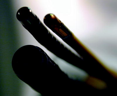

Fig. 3.3
Variations in the size of bronchoscopes
The difference between 1 mm in the working channel may not seem significant, but when you have an accessory tool that occupies 1.8 mm in a channel that is only 2.0 mm, there is no room to apply suction should your field of view become obscured, forcing you to remove the tool and start all over again. For the interventional pulmonologist, having the largest working channel can be a great friend and make for faster and safer procedures.
A Particular Function that the Bronchoscope May Provide
Over the past several years, there has been an explosion in technology development for the interventional pulmonologist. Much of this development is directed toward accessories placed through the working channel aimed at assisting the endoscopist in navigating through the airways, improving their diagnostic yield or providing better therapeutic options. Examples include electromagnetic navigation, alveoloscopy, and optical coherence tomography. Some of these technologies are still in their development stages and are not quite ready for clinical applications.
But there have also been developments to the bronchoscope itself. Newer technologies allow for integrated systems using the same bronchoscope to provide information using either different light frequencies to detect abnormal lung tissue (narrow band imaging, Fig. 3.4, or autofluorescence examination) and even the use of ultrasound technology to view through the airways themselves into the mediastinum to allow for sampling of these areas.
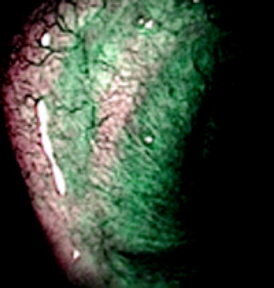

Fig. 3.4
Narrow band imaging
Efforts have been made to overcome the loss of suctioning ability when a large accessory tool has been placed in the working channel by designing scopes with a second working channel. But because of space limitations to the overall size of the bronchoscope, efforts to make this working channel large enough to be helpful have not been very successful.
Concerns over cross infection of patients undergoing bronchoscopy have prompted some manufacturers to develop bronchoscopes that can be steam sterilized and others that have a single use sterile sheath that can be placed over the entire scope (Fig. 3.5). The initial designs of these sheaths were cumbersome and very costly, but recent improvements seem very promising.
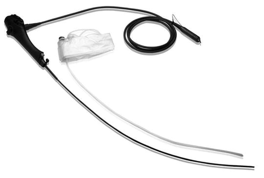

Fig. 3.5
Sterile sheathed bronchoscope
We are now just beginning to see high-definition bronchoscopes come to the market, and they are truly incredible. Because of the difference in size between bronchoscopes and gastroenterology scopes, it has taken a while longer for this technology to become available as our peers in gastroenterology have had this technology for several years. One must also remember that as the saying goes: “you are only as strong as the weakest link.” This also holds true to the world of high definition. You cannot expect to get the proper imaging from these scopes without proper high-definition monitoring, processing, and cabling.
As technologies emerge to assist the endoscopist in navigating to peripheral areas of the lung that are unable to be visualized directly, one would expect the manufacturers themselves to develop their own “extendable working channel” and not have to rely on add-on systems to provide this function.
This cascade of technology development over the past several years should continue if reimbursement can be established for these modalities. But, to date, there has not been much success in providing payment for any of these, and this certainly will affect future development.
Determining Selection
When considering the purchase of a bronchoscope, one needs to consider whether you are adding to your current inventory or beginning a new program. Even if starting fresh, you will no doubt have an inventory of some bronchoscopes that are being used by others performing bronchoscopy procedures that you will have access to. It is highly unlikely, in this economic environment, that the administration will grant a blank check and not consider the value of those older but still functional scopes. That being said, the IP program director needs to compile a list of the number, size, and type of scopes necessary to maintain a proper inventory. There are many factors that one must consider when determining how many scopes a program needs, including but not limited to:
1.
Current volume of procedures and growth over the first 3–5 years.
2.
Types of patients you see and plan on seeing. (This will determine the size and types of the scopes you will need to purchase.)
3.
Staffing patterns – This will determine the volume of patients you can handle.
4.
Current and future reprocessing methods – To determine turnaround time for the availability of scopes.
5.
Locations of procedures being performed – If procedures are performed in multiple locations at the same time, there may be less scopes available.
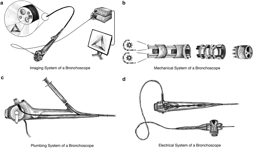
There are a variety of styles and sizes of bronchoscopes available for purchase. Most perform similar functions in that they allow the operator to visualize the conducting airways by using four integrated systems all contained in a plastic tube that is no more than a half inch in diameter.
1.
Image System – Responsible for transmitting the image from a lens at the end of the scope back to a video processor where the image is displayed on a monitor (Fig. 3.6a).
2.
Mechanical System – Allows navigation through the airways via a system of levers, wires, and hinged metal bands (Fig. 3.6b).
3.
Plumbing System – The working channel provides a communication between the upper end of the instrument and the distal end of the scope to allow for removal of blood or fluid to allow for proper viewing (Fig. 3.6c).
4.
Electrical System – Provides for a light system to illuminate the airway so that navigation can take place. Also allows for operator to perform functions using remote buttons on scope like image capture or changing light exposure to the tissue (Fig. 3.6d).

Fig. 3.6
Bronchoscopy systems
Proper Care of the Bronchoscope in an IP Program
Conventional Bronchoscopy Versus Interventional Pulmonology
Proper care of the bronchoscope and the resultant prevention of damage will have great financial implications as well as implications for the overall success of an interventional pulmonology program. Let us first consider the differences between an interventional and a conventional bronchoscopy program.
The work performed in conventional bronchoscopy is usually related more toward the diagnosis of disease and not the treatment or therapeutic intervention of that process. Much of the “diagnosis” consists of sampling the airway by washing, brushing, or removing small bits of tissue with a biopsy forceps. Even the use of needle aspiration for mediastinal lymph node biopsies is sometimes looked upon as advanced procedures by conventional bronchoscopists.
The accessory tools used by the interventional bronchoscopist carry with them a much higher risk of damage to the bronchoscope itself. Specialized training in the use of these tools in preventing damage to the bronchoscope is critical to the success of the interventional program.
Damage to any form of an endoscope can be costly, but damage to the bronchoscope can be especially so, for two reasons.
First, the size of the bronchoscope is limited to the size of the space it has to pass, so it is very difficult to navigate through the vocal cords and see much more than the central airways with a scope larger than 6–7 mm in diameter. Because of this size limitation, the working parts of the bronchoscope are much smaller and therefore more delicate than, for instance, a gastroscope.
The other reason for more costly repairs is the nature of the development of interventional pulmonology itself over the past several years. Academic programs have been graduating fellows at an increasing pace because of the demand by hospitals to develop their own interventional programs. Industry has been trying to keep pace with this demand by producing technologies to meet the demand of physicians for improved products as well as the development of new technologies. Because of these reasons, academic centers have been inundated with new equipment to pass through the bronchoscope. Much of this equipment is either very sharp or pass extreme heat, cold, or electrical current through them to perform their role in assisting in the diagnosis or treatment of diseased tissue.
Stay updated, free articles. Join our Telegram channel

Full access? Get Clinical Tree


