(1)
Ospedale Civile di Vigevano, Vigevano, Italy
Introduction
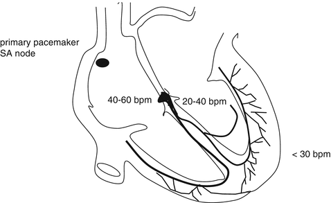
The excitation-conduction system of the heart is controlled by the sinoatrial (SA) node, which is located in the right atrium, near the junction between the atrium and the superior vena cava. The SA node emits rhythmic electrical impulses at a higher rate than the rest of the cardiac tissue, including the atrioventricular (AV) node, which fires at rates ranging from 40 to 60 beats per minute (bpm), and other segments of the conduction system and the myocardial tissue itself (20–40 bpm) (Fig. 5.1). As noted early, arrhythmias are anomalous cardiac rhythms caused by the abnormal formation and/or conduction of the electrical impulse. They can be broadly classified as tachyarrhythmias, characterized by rapid heart rates exceeding 100 bpm, and bradyarrhythmias, with slow rates of less than 60 bpm.


Fig. 5.1
The cardiac conduction system. The SA node is located in at the junction between the right atrium and the superior vena cava. The AV node is located in the right atrium near the coronary sinus. The bundle of His is located at the septal level. It divides to form the right and left bundle branches, which supply the right and left ventricles, respectively. The left bundle branch divides to form the anterosuperior and posteroinferior fascicles
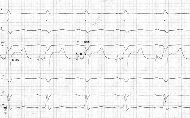
Useful insight into the bradyarrhythmias can be obtained with intracardiac ECG, which is performed with a catheter electrode placed inside the cardiac chambers. The tracing obtained with this technique (Fig. 5.2) includes three main polyphasic elements:


Fig. 5.2
Comparison of surface ECG tracings (leads I, II, III, aVF, V1, and V6) and intracardiac ECG (IC-ECG) tracing (4th from the top). The three main elements of the IC-ECG are seen: the A deflection (representing activation of the atrium); the H deflection (activation of the His bundle); and the V deflection (activation of the ventricular myocardium). See text for details on measurement of the PA, AH, and HV intervals. Paper speed: 100 mm/sec
D1 ——————- I
D2 ——————-II
D3 ——————-III
aVF —————– aVF
V1 ——————- V1
V6 ——————- V6
Endo —————- IC-ECG
the A deflection, which represents activation of the atria;
the H deflection, which corresponds to activation of the common trunk of the His bundle and has a normal duration of approximately 20–25 ms;
the V deflection, which reflects activation of the ventricular myocardium.
If the surface and intracardiac ECGs are recorded simultaneously, three important intervals can be measured:
The PA interval, which is measured from the onset of the P wave on the surface ECG to the first rapid A deflection. It represents the time required for the stimulus to travel from the SA node to the AV node (normally 15–25 ms).
The AH interval, which is measured from the onset of the A deflection to the onset of the H deflection. It reflects the conduction time through the AV node (normally 60–120 ms).
The HV interval, from the onset of the H deflection to the first rapid component of the V deflection, represents the time needed for depolarization of the His bundle, the bundle branches, and the Purkinje network (normally 35–55 ms).
These elements are necessary for subsequent understanding of the locations of atrioventricular blocks (AVBs) and their clinical significance and prognostic implications.
Classification and Electrocardiographic Characteristics
Several arrhythmia classification systems have been proposed at the Italian and international levels. Here, we will use the system proposed some years ago by the Italian Association of Hospital Cardiologists (Table 5.1).
Table 5.1
Italian association of hospital cardiologists classification of the arrhythmias (from Alboni et al., 1999)
Tachycardias | Bradycardias | Premature beats | |
|---|---|---|---|
Supraventricular | Ventricular | ||
Sinus tachycardia | Ventricular tachycardia (VT) | Sinus bradycardia | Supraventricular premature beats |
Atrial tachycardia | – nonsustained VT | Sinus arrhythmia | Ventricular premature beats |
Atrial flutter (AFL) | – sustained monomorphic VT | Sinoatrial blocks | |
– typical | – fascicular VT | Sinus arrest | |
– atypical | – outflow-tract VT | AV blocks (AVBs) | |
Atrial fibrillation (AF) | – polymorphous VT | – first degree | |
– Paroxysmal | – Torsade de pointes | – second degree | |
– Persistent | Ventricular fibrillation | • Wenckebach | |
Accelerated idioventricular rhythm (AIVR) | • Mobitz 2 | ||
– Permanent | • 2:1 | ||
• Advanced | |||
– third degree | |||
AV dissociation | |||
AV nodal reentrant tachycardia (AVNRT) | |||
– slow-fast | |||
– fast-slow | |||
– slow-slow | |||
Atrioventricular reentrant tachycardia (AVRT) caused by: | |||
– an orthodromic manifest (WPW) accessory pathway | |||
– an antidromic manifest (WPW) accessory pathway | |||
– a concealed accessory pathway | |||
– a concealed, slow-conduction accessory pathway (Coumel type) | |||
– nodoventricular or nodofascicular fibers (Mahaim type) | |||
Junctional ectopic tachycardia | |||
Arrhythmias Caused by Abnormal Impulse Formation
Healthy individuals at rest exhibit a sinus rhythm with rates ranging from 60 to 100 bpm. Variations can be caused by various factors related to the autonomic nervous system, the most common of which involves rate fluctuations related to the respiratory cycle (respiratory sinus arrhythmia).
Sinus Bradycardia
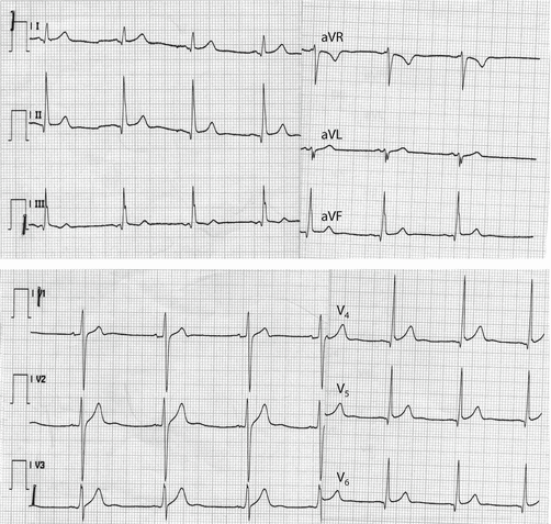
Sinus bradycardia is characterized by a regular rhythm and a rate of <60 bpm. It can be further classified as mild (rates between 50 and 59 bpm), moderate (40–49 bpm), or severe (<39 bpm) (Figs. 5.3 and 5.4). It is important to recall that in young, healthy subjects, nocturnal sinus rhythms at rates of 35–40 bpm are considered normal.


