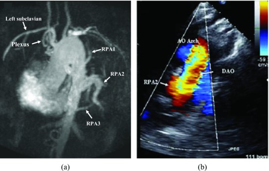
Figure 48.2 MRI image show that there are three right pulmonary arteries (RPA 1, 2, 3) derived from descending aorta, and a left sided plexus of arteries (plexus) derived from left subclavian artery (A). Transthoracic super sternal view shows aortic arch and one right pulmonary artery derived from descending aorta (located in right side) (B). These two images are comparable.

Magnetic resonance imaging showed: there are three right pulmonary arteries (RPA 1, 2, 3) derived from descending aorta, and a left sidedplexus of arteries (plexus) derived from left subclavian artery. The left lung is smaller than right due to short of blood supply (Figure 48.3). A hypoplasia of left lung and plexus of vessels which originates from the left subclavian and supplies collaterals to the arterial circulation of the left lung, and demonstrated those findings by echocardiography (Figure 48.2
Stay updated, free articles. Join our Telegram channel

Full access? Get Clinical Tree


