Year
Author
Type
Configuration
N
Duration
Patency rate
Distribution
Systemic anticoagulation
1971
Eger
PVC tube
T-shaped
36
ND
ND
ULAV
Yes
1979
Majeski
Javid
Looped
2
210 min
100 % (2/2)
ULAV
ND
1981
Nunley
Sundt
Straight
8
ND
100 % (8/8)
ULAV
ND
Ventriculoperitoneal shunts
Straight
1984
Eggink
ND
T-shaped
ND
ND
ND
ND
ND
1986
Khalil
Javid
Straight
5
2–6 h
100 % (5/5)
LEAV
No
Balloon-type shunts
Straight
Nasogastric tube
Straight
1986
Nichols
Javid and Sundt
Looped
13
127 min
100 % (13/13)
ULAV
No
1989
Johansen
PVC intravenous tube
Looped
1
16 h
100 % (1/1)
LEAV
No
1992
Husain
PVC Suction catheter
ND
5
3–6 h
100 % (5/5)
LEA
No
1995
Reilly
Javid
Straight
1
36 h
100 % (1/1)
SMA
No
1996
Starr
ND
ND
3
ND
100 % (3/3)
LEA
No
1999
Reber
Heyer-Schulte carotid shunt
T-shaped
7
90–390 min
100 % (7/7)
ULAV
No
2000
Granchi
Argyle
Straight
19
47–3,130 min
100 % (19/19)
ULAV
No
2002
Sriussadaporn
PVC intravenous and extension tube
Looped
7
60–180 min
100 % (7/7)
LEAV
No
2003
Parry
Argyle
Straight
18
22 h
100 % (18/18)
LEA
No
thoracostomy tube
Straight
Pruitt-Inahara
T-shaped
2003
Rozycki
Chest tube
Straight
13
24–54 h
83 % (15/18)
ULAV
No
Angiocatheter
Straight
Argyle
Straight
Pruitt-Inahara
T-shaped
2004
Hossny
PVC Suction catheter
Straight
7
ND
100 % (7/7)
LEAV
No
2004
Nalbandian
ND
Looped
1
24 h
100 % (1/1)
LEAV
No
2006
Rasmussen
Javid
Straight
30
2 h
86 %a(19/22)
ULAV
No
Argyle
Straight
12 %b(1/8)
2006
Chambers
Javid and Sundt
Straight
20
<6 h
78 % (21/27)
ULAV
No
Commercially available shunts (i.e., Javid, Sundt, Argyle, and Pruitt-Inahara carotid shunts) are first designed to provide temporary carotid bypasses for cerebral circulation during carotid endarterectomy procedures, and now they are widely used as an adjunct to DCS in severe vascular injuries.
The Sundt shunt (internal and external) is constructed of silicone elastomer with stainless steel spring reinforcement to minimize kinking and occlusion of the cannula lumen and to aid in the ease of insertion of the proximal and distal ends [17]. The ends of the shunt have cone-shaped bulbs to facilitate fixation of the shunt in the vessel. The bulb fits so firmly against the wall of the vessel that there is no bleeding around the distal bulb.
The Javid shunt has fusiform swellings at each end and is held in place by external clamps [18]. It is most commonly used in the superficial femoral artery and vein [19]. Argyle shunts are usually used in the tibial and brachial arteries. Both the Argyle and Javid are in-line shunts, while the few Sundt shunts are either in-line or looped. However, in the military experiences in Iraq, Rasmussen et al. [19] did not see any differences in patency rates among the Javid, Sundt, and Argyle shunts, although the patient number was small.
The Pruitt-Inahara shunt is the same length as the Javid, but is held in place by incorporated inflatable balloons at each end. They are dual-lumen devices with balloons at both the distal and proximal ends of the shunt with a T-piece. The balloons, when inflated independently, act as a stabilization mechanism to maintain the position of the shunt when it is placed within the arteries. An external safety balloon located on the inflation arm leading to the distal balloon acts as a mechanism to relieve pressure on the distal balloon in the event it inflates above maximum stated volume. The external safety balloon feature reduces the possibility of balloon overinflation and resultant vessel damage [18, 20]. The Pruitt-Inahara carotid shunt instantly verifies pulsatile flow, and an arteriogram can be performed to verify flow to the hand or foot. Its occlusion balloons eliminate the need for clamps, thereby reducing vessel trauma, using a small incision, minimal dissection, and a short arteriotomy [21].
Self-made shunts such as the endotracheal suction catheter [13, 22], nasogastric tube [15], ventriculoperitoneal shunt [23], thoracostomy tube [21, 24], and angiocatheter [24] are readily available in various sizes, inexpensive, and non-toxic. The Indian surgeon Husain et al. [13] reported the inexpensive and easily available polyvinylchloride (PVC) disposable endotracheal suction catheters, which were successfully used as temporary intravascular shunts in five popliteal artery trauma patients during the delay between the time of injury or presentation and the vascular repair. The shunts provided good outcomes with all limbs viable without complications.
Choosing the right shunt diameter size is critical. However, the surgeon selects the shunt size mostly based on clinical experience and estimation of vessel size [25]. One principle is that it is essential to never have an oversized shunt in order to minimize endothelium dysfunction. Many authors prefer to loop the shunt while securing it in vessel. However, Chambers et al. [6] suggested to avoid looping of the shunts to help minimize both the resistance to flow and likelihood of dislodgment as both complications occurred intraoperatively early in their military experience. They advocated ensuring the shunt lies straight; they needed a significant length to lie within the vessel lumen above and below the site of injury.
How Long Can the Shunt Stay Patent?
The success rate of arterial revascularization is inversely proportional to the duration of the ischemic time. According to the histological studies of Malan [26], the critical warm ischemic time for striated muscle is 6–8 h. Blood flow must be reestablished within this time to avoid irreversible muscle and nerve damage. The presence of soft tissue loss, disruption of arterial and venous collateral vessels, or hypotension may further decrease the length of this critical period. After vascular shunts have been inserted, potentially life-threatening injuries to the head, chest, or abdomen can be diagnosed and treated in a more orderly fashion. Similarly, use of these vascular shunts can prevent the need for the amputation of potentially salvageable extremities because of the prolonged ischemic time during other operative procedures. Therefore, the amount of time the intravascular shunt can maintain patency is of importance to the trauma surgeon.
The reported shunt times in the literature ranged from a couple of minutes to as long as several days. The longest shunt time of up to 10 days occurred in a civilian case (David V. Feliciano, Atlanta, GA, 2009 personal communication). But the longest dwell time recorded in the literature was reported by Granchi [25], who demonstrated that shunts could be left in place and maintained distal perfusion for up to 52 h without systemic anticoagulation in complex extremity injuries. Although Rozycki et al. [24] reported a dwell time of 54 h for a Pruitt-Inahara shunt left in the brachial artery, this shunt was occluded at reoperation, resulting amputation in the end. Intravascular shunts have been used to maintain the vascular patency of the extremities, with times ranging from 12 to 17 h in three patients undergoing air transfer to a level I trauma center for definitive care [27]. No evidence of shunt thrombosis or distal emboli was observed. However, Rasmussen et al. [19] argued that in war time maintaining the shunt patency for 2 h is sufficient because of fast evacuation patterns and relatively fewer shunt-related complications.
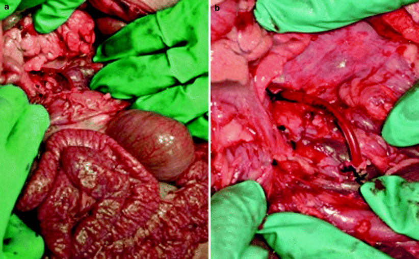
Experimentally, Ding compared four different shunt indwelling times in previously established superior mesenteric artery (SMA) injury models [28] and aimed to determine the safest duration in the setting of DCS. Their study suggested that TIVS can be used as an important DCS adjunct to maintain patency safely for 6 h (Fig 18.1); however, prolonged duration of an indwelling vascular shunt (over 9 h) caused endothelial injury in such SMA injuries in the setting of DCS [29]. Their recent study [30] showed that prolonged indwelling time of temporary vascular shunts is associated with increased endothelial injury, and, when possible, vascular reconstruction following the use of shunts should include an interposition graft after debridement of the arterial edges that interfaced with the shunt. Finally, to minimize intimal injury to the native vessel, this model suggests that the indwell times of shunts should be <9 h.
 < div class='tao-gold-member'>


Only gold members can continue reading. Log In or Register to continue
Stay updated, free articles. Join our Telegram channel
Full access? Get Clinical Tree
