Fig. 27.1
HeartMate 3 outflow-graft anastomosis on the ascending aorta by adoption of a side clamp (Illustration by Ilaria Bondi’s Peppermint Advertising)
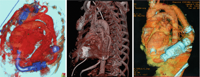
Additionally, the outflow may be anastomosed to the descending aorta through a left thoracotomy (◘ Fig. 27.2) as suggested by such manufactures as Jarvik Inc. (Jarvik Heart, NY) for Jarvik 2000 LVAD placement [7, 8]. Or alternatively the descending aorta may be the site for any other LVAD outflow-graft anastomosis in case of redo operation implantation or in case of bridge-to-heart transplantation (Htx) intention to avoid any eventual risky sternal reentry and provide a technically easier surgery.


Fig. 27.2
Descending aorta outflow-graft placement by 3D computed tomography (CT) scan reconstruction
Outflow-graft protection is of major interest in the bridge-to-transplant therapy with left ventricular assist devices (LVADs). Avoiding adhesions of the outflow graft to the sternum prevents possible graft damages and bleeding events while performing re-thoracotomy for Htx or pump exchange. Furthermore, it reduces the time used for preparation of the anatomical structures before Htx.
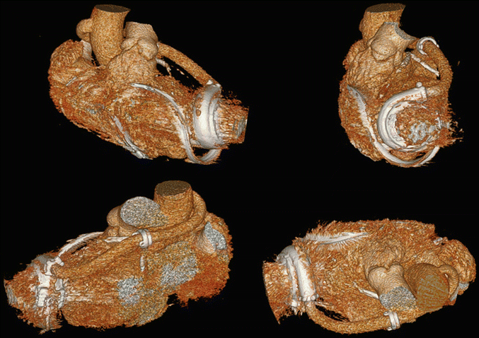
A novel surgical approach for outflow LVAD placement may be its positioning through the transverse sinus (◘ Fig. 27.3), thus performing the anastomosis, after its adequate trimming and orientation, on the right posterolateral side of the ascending aorta [9]. The positioning of the outflow graft plays an important role in the long-term outcome of the patient. False positioning leads to turbulences in the blood flow, resulting in a high risk of outflow-graft thrombosis or kinking. The flow in the outflow graft seems not to be affected by tunneling the outflow graft through the transverse sinus. Due to the augmented physiological angle of the anastomosis of the outflow graft to the aorta, the accelerated blood flow in the ascending aorta is physiological and aortic insufficiency might be decreased. Preparing the transverse sinus for outflow-graft tunneling is not time-consuming, and, with surgical training, the additional amount of time used for this technique compared to the standard procedure is reduced to a minimal difference.


Fig. 27.3
HVAD outflow graft tunneling through the transverse sinus by 3D computed tomography (CT) scan reconstruction
Moreover, in case of redo scenarios and severely calcified aorta, the outflow may be sutured on the axillary artery [10] (◘ Fig. 27.4). The left ventricular apex is accessed through a left mini-thoracotomy. The outflow graft is tunneled through a small incision in the fourth intercostal space and then subcutaneously to the subclavian region. In order to avoid any outflow-graft kinking and compression at the passage between the ribs, a covering vascular prosthesis with a ring-reinforced Gore-Tex graft may be adopted. After division of the left axillary artery, an end-to-end anastomosis is performed to the proximal part, and the distal vessel is connected end-to-side through a fenestration in the outflow graft. This technique may achieve a more direct blood flow into the aorta and reduces cerebrovascular events while avoiding excessive flow to the arm. Other authors [10] suggest simply a single end-to-side anastomosis between the outflow graft and axillary artery. A distal banding of subclavian artery may be considered to avoid hyperflow and postoperative edema in left arm. The only significant potential adverse event may be compression of outflow graft between under the clavicle, particularly when the left arm is elevated > 90°.


