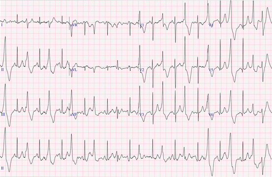
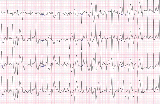
Fig. 5.1
Baseline 12-lead ECG showing sinus bradycardia
An exercise test was performed, and she exercised for 11 min and 59 s on the Bruce protocol. Her resting heart rate before starting the test was 55 beats per minute rising to a maximum of 173 beats per minute during peak exercise. There was an appropriate blood pressure response to exercise reaching 165/67 and returning to baseline upon rest. At the start of the test her ECG showed normal sinus rhythm but on exertion once her heart rate reached 140 bpm frequent ventricular ectopics began to appear. At 150 bpm ventricular couplets were seen and as her heart rate rose further these became more frequent and bi-directional in nature. At that point the test was terminated (Figs. 5.2 and 5.3).


Fig. 5.2
Frequent ventricular ectopic beats during exercise ECG

Fig. 5.3
Frequent ventricular couplets of bidirectional nature during exercise ECG
A diagnosis of catecholaminergic polymorphic ventricular tachycardia (CPVT) was made and she was prescribed atenolol. Since the introduction of medical therapy repeat exercise testing has shown a reduced amount of ventricular ectopy on exercise and she has had no further episodes of collapse.
Case Description 2
An 8-year-old boy who had been living with his grandparents abroad moved to the UK to live with his parents. Three months later, the boy had gone to bed as usual when his mother heard a scream coming from his room. When she went to investigate she found him collapsed on the floor. She called for an ambulance and started CPR. The first responder arrived after 5 min and found the boy to be in pulseless electrical activity; he continued basic life support. When the paramedic crew arrived the boy was intubated, resuscitation was continued, and by 17 min cardiac output had returned. The boy was air-lifted to the children’s hospital for further management.
On arrival in the emergency department the boy had a heart rate of 140–190 bpm, showing a variable rhythm with runs of monomorphic and polymorphic ventricular tachycardia (Fig. 5.4), supraventricular tachycardia, bigemeny and trigemeny. The rhythm changes were noticed to be associated with the painful stimuli such as suction and blood sampling. The boy tolerated the rhythm changes well with no further loss of cardiac output. He was transferred to the intensive care unit where he was cooled for 24 h. During this time his electrolytes were optimized and he was started on lidocaine and esmolol infusions. After 24 h he was re-warmed, sedation was reduced, and he was successfully extubated. The esmolol infusion was changed to oral propranolol and other infusions discontinued. He went on to make a good recovery, and was discharged with no evidence of neurological injury. He has continued to do well and is currently managed on oral flecanide and propranolol.


Fig. 5.4
Rhythm strip in A&E showing ventricular ectopy and runs of ventricular tachycardia
< div class='tao-gold-member'>
Only gold members can continue reading. Log In or Register to continue
Stay updated, free articles. Join our Telegram channel

Full access? Get Clinical Tree


